Difference between revisions of "20.109(S20):Treat cells with etoposide and analyze RNA-seq data (Day2)"
Noreen Lyell (Talk | contribs) (→Navigation links) |
Becky Meyer (Talk | contribs) (→Part 2: Treat cells with etoposide) |
||
| (10 intermediate revisions by 3 users not shown) | |||
| Line 6: | Line 6: | ||
In this module, you will assess gene expression using two methods: quantitative PCR (qPCR) and RNA-seq. For timing reasons you will not be able to prepare and submit your treated samples for RNA-seq. Instead you will use RNA-seq data generated by the teaching faculty. You will, however, complete the same procedure used by the teaching faculty to prepare the RNA samples and then use your samples in a qPCR experiment to confirm the RNA-seq results. | In this module, you will assess gene expression using two methods: quantitative PCR (qPCR) and RNA-seq. For timing reasons you will not be able to prepare and submit your treated samples for RNA-seq. Instead you will use RNA-seq data generated by the teaching faculty. You will, however, complete the same procedure used by the teaching faculty to prepare the RNA samples and then use your samples in a qPCR experiment to confirm the RNA-seq results. | ||
| − | To begin your assay, you will first induce DNA damage by incubating your cells with etoposide. Etoposide is a chemotherapy drug that generates DNA strand breaks by forming a ternary complex with DNA and topoisomerase II. This prevents religation of the DNA strands after unwinding. This effects cancer cells more than non-cancerous cells because cancer cells divide much faster. We will use etoposide to damage the DNA then examine the | + | To begin your assay, you will first induce DNA damage by incubating your cells with etoposide. Etoposide is a chemotherapy drug that generates DNA strand breaks by forming a ternary complex with DNA and topoisomerase II. This prevents religation of the DNA strands after unwinding. This effects cancer cells more than non-cancerous cells because cancer cells divide much faster. We will use etoposide to damage the DNA then examine the transcriptome of DLD-1. |
| − | + | ||
| − | + | ||
==Protocols== | ==Protocols== | ||
| − | ===Part 1: | + | ===Part 1: BE Communication Lab workshop=== |
| − | + | Our communication instructors, Dr. Sean Clarke and Dr. Prerna Bhargava, will join us today for a workshop on preparing and organizing your Journal Club presentations. | |
| − | + | ===Part 2: Treat cells with etoposide=== | |
| + | In this exercise, you will induce DNA damage in cells that were seeded by the teaching faculty (using the cells you seeded during the previous laboratory session). Next class you will purify RNA from your cells for quantitative PCR analysis. Control, no treatment flasks will be prepared by instructors. | ||
#Prepare your working space within the tissue culture hood. | #Prepare your working space within the tissue culture hood. | ||
| Line 22: | Line 21: | ||
#*Determine the volume of etoposide stock (100 mM) you need to add to the media for a final concentration of 100 μM in the 10 mL aliquot. | #*Determine the volume of etoposide stock (100 mM) you need to add to the media for a final concentration of 100 μM in the 10 mL aliquot. | ||
#*Prepare your media +etoposide. | #*Prepare your media +etoposide. | ||
| − | #Retrieve | + | #Retrieve one T75 flask of DLD-1 cells from the 37 °C incubator and visually inspect your cells with a microscope. |
#*Record your observations concerning media color, confluency, etc. in your laboratory notebook. | #*Record your observations concerning media color, confluency, etc. in your laboratory notebook. | ||
| − | #Move your | + | #Move your flask into the tissue culture hood. |
| − | #Aspirate the spent media from | + | #Aspirate the spent media from the flask. |
| − | + | #Add 5 mL of PBS to the flask and rock the plate gently to wash the cells. | |
| − | #Add 5 mL of PBS to | + | |
#Aspirate the PBS from each flask. | #Aspirate the PBS from each flask. | ||
| − | + | #Add 10 mL of the media containing etoposide that you prepared in Step #2 to the flask. | |
| − | #Add | + | #Carefully move the 'etoposide treated' cells to the 37 °C incubator for 60 min. |
| − | #Carefully move the 'etoposide treated' | + | #When you are done, return to the main laboratory space. |
| − | # | + | #When the 60 min incubation is complete, return to the tissue culture space and retrieve your flask from the 37 °C incubator. |
| − | # | + | #Aspirate the media containing etoposide from the flask. |
| − | #Aspirate the media containing etoposide from | + | #Add 10 mL of fresh media to the flask. |
| − | + | #Label the flask 'DLD-1 +etop' to denote that the cells were treated with etoposide. | |
| − | #Add 10 mL of fresh media to | + | #*Move the flasks to the 37 °C incubator. |
| − | #Label the | + | #Clean the hood and return the main laboratory space. |
| − | #*Move | + | |
| − | # | + | |
| − | + | ||
| − | + | ||
| − | + | ||
| − | + | ||
| − | + | ||
| − | + | ||
| − | + | ||
| − | + | ||
| − | + | ||
| − | + | ||
| − | + | ||
| − | + | ||
| − | + | ||
| − | + | ||
| − | + | ||
| − | + | ||
| − | + | ||
| − | + | ||
| − | + | ||
| − | + | ||
| − | + | ||
| − | + | ||
| − | + | ||
| − | + | ===Part 3: Analyze RNA-seq data=== | |
| + | Today you will analyze the RNA-seq data gathered from untreated DLD-1 and etoposide-treated DLD-1 cells. Following RNA purification, the samples were submitted to the BioMicro Center for Illumina sequencing. Illumina sequencing technology, or sequencing by synthesis (SBS), is used for massively parallel sequencing with a proprietary method that detects single bases as they are incorporated into growing DNA strands. | ||
| − | + | Complete Exercise #2 developed by Amanda Kedaigle, Anne Shen & Prof. Ernest Fraenkel. | |
| − | + | ||
| − | + | ||
| − | + | ||
| − | + | ||
==Reagents== | ==Reagents== | ||
Latest revision as of 19:56, 10 March 2020
Contents
Introduction
In this module, you will assess gene expression using two methods: quantitative PCR (qPCR) and RNA-seq. For timing reasons you will not be able to prepare and submit your treated samples for RNA-seq. Instead you will use RNA-seq data generated by the teaching faculty. You will, however, complete the same procedure used by the teaching faculty to prepare the RNA samples and then use your samples in a qPCR experiment to confirm the RNA-seq results.
To begin your assay, you will first induce DNA damage by incubating your cells with etoposide. Etoposide is a chemotherapy drug that generates DNA strand breaks by forming a ternary complex with DNA and topoisomerase II. This prevents religation of the DNA strands after unwinding. This effects cancer cells more than non-cancerous cells because cancer cells divide much faster. We will use etoposide to damage the DNA then examine the transcriptome of DLD-1.
Protocols
Part 1: BE Communication Lab workshop
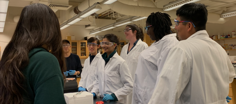
Our communication instructors, Dr. Sean Clarke and Dr. Prerna Bhargava, will join us today for a workshop on preparing and organizing your Journal Club presentations.
Part 2: Treat cells with etoposide
In this exercise, you will induce DNA damage in cells that were seeded by the teaching faculty (using the cells you seeded during the previous laboratory session). Next class you will purify RNA from your cells for quantitative PCR analysis. Control, no treatment flasks will be prepared by instructors.
- Prepare your working space within the tissue culture hood.
- Calculate the volume of etoposide stock needed for DNA damage induction.
- Obtain an aliquot of pre-warmed media from the 37 °C water bath.
- Transfer 10 mL of the media into a 15 mL conical tube.
- Determine the volume of etoposide stock (100 mM) you need to add to the media for a final concentration of 100 μM in the 10 mL aliquot.
- Prepare your media +etoposide.
- Retrieve one T75 flask of DLD-1 cells from the 37 °C incubator and visually inspect your cells with a microscope.
- Record your observations concerning media color, confluency, etc. in your laboratory notebook.
- Move your flask into the tissue culture hood.
- Aspirate the spent media from the flask.
- Add 5 mL of PBS to the flask and rock the plate gently to wash the cells.
- Aspirate the PBS from each flask.
- Add 10 mL of the media containing etoposide that you prepared in Step #2 to the flask.
- Carefully move the 'etoposide treated' cells to the 37 °C incubator for 60 min.
- When you are done, return to the main laboratory space.
- When the 60 min incubation is complete, return to the tissue culture space and retrieve your flask from the 37 °C incubator.
- Aspirate the media containing etoposide from the flask.
- Add 10 mL of fresh media to the flask.
- Label the flask 'DLD-1 +etop' to denote that the cells were treated with etoposide.
- Move the flasks to the 37 °C incubator.
- Clean the hood and return the main laboratory space.
Part 3: Analyze RNA-seq data
Today you will analyze the RNA-seq data gathered from untreated DLD-1 and etoposide-treated DLD-1 cells. Following RNA purification, the samples were submitted to the BioMicro Center for Illumina sequencing. Illumina sequencing technology, or sequencing by synthesis (SBS), is used for massively parallel sequencing with a proprietary method that detects single bases as they are incorporated into growing DNA strands.
Complete Exercise #2 developed by Amanda Kedaigle, Anne Shen & Prof. Ernest Fraenkel.
Reagents
- etoposide, stock = 100 mM (Sigma-Aldrich)
Next day: Purify RNA from etoposide-treated cells and generate cDNA
