20.109(F19):Module 1
Contents
Module 1
Lecturer: Bevin Engelward
Instructors: Noreen Lyell, Leslie McClain and Becky Meyer
TAs: Colin Kim and Shelbi Parker
Lab manager: Hsinhwa Lee
Overview
Cancer is a disease of the genome. Cancer is caused by accumulated mutations in genes that lead to phenotypic advantages in the progression from normal to metastatic cancer. While we know a lot about the kinds of changes that cancer cells undergo, we do not yet fully understand where mutations come from in the first place. What is clear is that changes to the DNA structure (e.g., DNA damage) can cause carcinogenic mutations. Given that DNA damage causes mutations that drive cancer progression, it is important to have effective tools for measuring DNA damage. In this module, you will learn about two different approaches for quantifying DNA damage. The first method relies on physical changes to the DNA structure that impact its ability to migrate when electrophoresed. This is a direct measure of DNA damage. The second method relies on antibody recognition of changes to proteins that occur as a result of cell signaling that is triggered by DNA damage. This is an indirect measure of DNA damage. Given the potentially deadly consequences of DNA damage, it is a good thing that our cells have robust ways to repair their DNA. Being able to measure DNA damage means that we can study DNA repair, which turns out to be a very important variable when it comes to why some people get cancer, and others do not.
In addition to learning many fundamental biological and engineering concepts, you will of course learn many laboratory techniques. In particular, you will learn about electrophoresis, mammalian cell culture, quantitative image analysis techniques, basic statistics, enzyme kinetics, molecular pathway analysis, impact of genetic factors, epigenetic analysis, antibody labeling and much more.
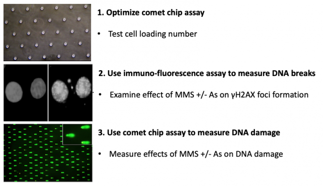
In terms of the specific experiments that you will do, in this module you will measure genomic instability using two techniques: a sub-nuclear foci assay (γH2AX immunofluorescence) and a microwell array (CometChip). You will use these methods to assess the effect of exposure to contaminants known to cause DNA damage. Specifically, you will assess the levels of DNA damage caused by low concentrations of methyl methanosulfate (MMS) and arsenite (As) alone and in combination.
Lab links: day by day
M1D1: Practice cell culture and begin gamma-H2AX assay
M1D2: Treat cells for gamma-H2AX assay and prepare CometChip for loading experiment
M1D3: Complete gamma-H2AX assay and perform loading experiment
M1D4: Load cells into CometChip and apply treatments for DNA damage experiment
M1D5: Complete DNA damage experiment
M1D6: Image CometChip
M1D7: Practice statistical analysis methods and complete data analysis
Assignments
Data summary
Mini-presentation
References
CometChip: A high-throughput 96-well platform for measuring DNA damage in microarrayed human cells. Journal of Visualized Experiments. (2014) 92: 1-11.
- A video of the procedure is linked here.
CometChip: Single-cell microarray for high-throughput detection of DNA damage. Methods in Cell Biology. (2012) 112: 247-268.

