20.109(F19):Begin Western blot analysis (Day3)
Contents
Introduction
Electrophoresis is a technique that separates large molecules by size using an applied electrical field and a sieving matrix. DNA, RNA and proteins are the molecules most often studied with this technique; agarose and acrylamide gels are the two most common sieves. The molecules to be separated enter the matrix through a well at one end and are pulled through the matrix when a current is applied across it. The larger molecules get entwined in the matrix and are stalled; the smaller molecules wind through the matrix more easily and travel farther away from the well. The distance a nucleic acid or amino acid fragment travels is inversely proportional to the log of its length. Over time fragments of similar length accumulate into “bands” in the gel.
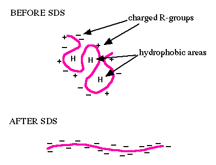
You will use sodium dodecyl sulfate-polyacrylamide gel electrophoresis (SDS-PAGE) to evaluate your purified protein. SDS is an ionic surfactant (or detergent), which denatures the proteins and coats them with a negative charge. Since denatured proteins are linear, they will move through the gel at a speed inversely proportional to their molecular weight, just like DNA on agarose gels. (Non-denatured proteins run according to their molecular weight, shape, and charge.) You will run a reference ladder containing proteins of known molecular weight and amount to determine the size and concentration of your purified FKBP12 protein. When running uninduced and induced samples side-by-side, you should see a protein band at the expected molecular weight for FKBP12 (~14 kDa + 3 kDa from the His-tag = 17 kDa), which may be very faint or non-existent in the uninduced control sample, but bright and thick in the induced sample. To visualize all the proteins released by the bacteria, you will stain the gels with Coomassie Brilliant Blue (actually, a variant called BioSafe Coomassie). Because this is a non-specific stain for all proteins, it will provide information concerning the purity of your protein sample.For Western blot analysis, a high quality antibody can have a relatively low affinity for its target protein. This is because the target is localized and concentrated on a blot, allowing the antibody to bind using both antibody “arms” thereby strengthening the association. Even an antibody that is loosely bound to the blot under these circumstances may dissociate then re-associate quickly since the local concentration of the target protein is high. The lower limit for protein detection is approximately 1 ng/lane, a value that varies with the size of the protein to be detected and the Western blotting apparatus that is used. For most polyacrylamide gels, the protein capacity for each lane is 100 to 200 μg (that would be 20 μL of a 5-10 μg/μL protein preparation). Thus, 1 ng represents a protein that is approximately 0.0005-0.001% of the total cellular protein (1 ng out of 100,000-200,000 ng). Proteins that make up a more significant fraction of the total protein population will be easier to detect.
Protocols
Part 1: Lyse cells
Part 2: Electrophorese cellular proteins
During the previous laboratory session, you reserved an aliquot of your induced and the uninduced cell lysate. In addition, the flow-through from the wash steps was stored. Today you will use SDS-PAGE to visualize the effectiveness of IPTG induction and the purification procedure.
- Retrieve the 15 μL aliquots of your induced and the uninduced cell lysates you prepared during the previous laboratory session. In addition, collect the flow-through from your wash steps and your purified, dialyzed protein solution.
- Transfer 25 μL from each of the wash flow-through samples into labeled 1.5 mL eppendorf tubes.
- Add 5 μL of Laemmli sample buffer to all of the samples prepared for SDS-PAGE
- This should include the uninduced / induced cell lysates, wash flowthroughs, elutions (from Part #2) and concentrated protein (from Part #2).
- Boil all samples for 5 min in the water bath located in the chemical fume hood.
- Secure the caps with the cap-locks located in the fume hood to ensure that the eppendorf caps do not pop open during the boiling step as this will result in your sample escaping the tube.
- If there is significant condensation at the lid of your tubes after boiling spin your samples before loading.
- You will load all samples and two molecular weight standards.
- A pre-stained ladder will be used to track the migration of your samples through the polyacrylamide gel.
- An unstained ladder with bands of known amounts of protein will be used to estimate protein concentration in your samples.
- Record the order in which you will load your samples and molecular weight standards in the polyacrylamide gel.
- When you are ready to load your samples, alert the teaching faculty.
- Please watch the demonstration closely to ensure your samples are correctly loaded and the polyacrylamide gel is not damaged during loading.
- Your samples will be electrophoresed at 200 V for 30-45 min.
- Following electrophoresis, use the spatula to carefully pry apart the plates that encase your polyacrylamide gel.
- Using wet gloves, transfer your polyacrylamide gel to a staining box and add enough dH2O to cover the gel.
- Wash the gel for 5 min at room temperature on the rotating table.
- Empty the water from the staining box in the sink.
- Be careful that the gel does not fall into the sink!
- Repeat Steps #12-13 a total of 3 times.
- Add 50 mL of BioSafe Coomassie to the staining box and incubate for 60 min at room temperature on the rotating table(or overnight depending on time).
- Empty the BioSafe Coomassie into the appropriate waste container in the chemical fume hood.
- Be careful that the gel does not fall into the waste container!
- Add 200 mL of dH2O to the staining box.
- Wash the gel for the remainder of the class on the rotating table.
- Replace the dH2O every 30 min.
Tomorrow the teaching faculty will transfer your gel to fresh dH2O and take a photograph. The image will be posted to the Class Data page.
Part 3: Transfer proteins to membrane
Reagents list
Next day: Complete antibody staining for Western blot analysis

