Difference between revisions of "Geometrical optics and ray tracing"
| Line 6: | Line 6: | ||
Light is an extraordinarily complicated, three dimensional, time dependent phenomenon. Completely describing a light field requires a function that quantifies the intensity, phase, wavelength, and propagation direction of light at every point in space. Needless to say, it can be difficult to conceptualize a light field. Fortunately, there is a simplified model of light that applies in situations where objects that interact with the light field are much larger than the wavelength of the light. | Light is an extraordinarily complicated, three dimensional, time dependent phenomenon. Completely describing a light field requires a function that quantifies the intensity, phase, wavelength, and propagation direction of light at every point in space. Needless to say, it can be difficult to conceptualize a light field. Fortunately, there is a simplified model of light that applies in situations where objects that interact with the light field are much larger than the wavelength of the light. | ||
| − | The simplified model is called ray optics. Ray optics abstracts the light field as a collection of lines pointing in the direction of propagation. | + | The simplified model is called ray optics. Ray optics abstracts the light field as a collection of lines pointing in the direction of propagation. The simplifications of ray optics offer a very good way to understand the function of most systems of lenses, mirrors, filters, and illumination sources. In many cases, ray optics can elucidate the function of a complicated optical system with just a few lines, triangles, and simple rules. |
The simplicity of ray optics comes at a cost. Ray optics glosses over important details like diffraction and interference. Thus, ray optics alone does not provide insight into detailed performance characteristics like resolution. | The simplicity of ray optics comes at a cost. Ray optics glosses over important details like diffraction and interference. Thus, ray optics alone does not provide insight into detailed performance characteristics like resolution. | ||
Revision as of 04:52, 4 February 2015
Light is an extraordinarily complicated, three dimensional, time dependent phenomenon. Completely describing a light field requires a function that quantifies the intensity, phase, wavelength, and propagation direction of light at every point in space. Needless to say, it can be difficult to conceptualize a light field. Fortunately, there is a simplified model of light that applies in situations where objects that interact with the light field are much larger than the wavelength of the light.
The simplified model is called ray optics. Ray optics abstracts the light field as a collection of lines pointing in the direction of propagation. The simplifications of ray optics offer a very good way to understand the function of most systems of lenses, mirrors, filters, and illumination sources. In many cases, ray optics can elucidate the function of a complicated optical system with just a few lines, triangles, and simple rules.
The simplicity of ray optics comes at a cost. Ray optics glosses over important details like diffraction and interference. Thus, ray optics alone does not provide insight into detailed performance characteristics like resolution.
This page introduces ray optics and discusses distortions called optical aberrations that occur in situations where one or more of the simplifying assumptions fail. Diffraction and resolution are discussed on a separate page.
Reflection and refraction
When incident light encounters an interface between two optical media, it can be
- reflected,
- scattered in a random direction,
- refracted,
- absorbed, or
- some combination of the above.
The systems of optical components that we will consider in 20.309 have smooth, non-absorbent surfaces. Their behavior is predominantly governed by reflection and refraction, so we won't spend a lot of time on scattering and absorption for now.
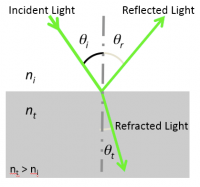
The law of reflection is pretty simple: states that the angle θr of a reflected is equal to the angle θi of the incident ray. (Angles are measured relative to a vector normal to the surface.) Incident and reflected rays are in the same plane.
Index of refraction
- The index of refraction $ n $ of a transparent optical medium is defined as the ratio of the speed of light in vacuum, $ c $, by the speed of light in the medium, $ v $.
- $ n = c / v $
Snell's law of refraction
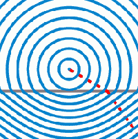
- Light incident at an interface between two media of distinct indices of refraction will be partly reflected back into the first medium and partly transmitted to the second medium, or refracted according to the Snell-Descartes law:
- $ n_i\ \sin \theta_i = n_t\ \sin \theta_t $
- with θ the angle measured from the normal of the boundary, $ n $ the refractive index (which has no units) of the medium, and the subscripts $ i $ and $ t $ referring to the incident and transmitted light, respectively.
- Snell's law implies that light passing from a medium of lower index to a higher index of refraction ($ n_t > n_i $) bends toward the surface normal ($ \theta_t < \theta_i $), whereas light traveling from a higher index to a lower index of refraction bends away from the normal. Remember, a higher index means a lower velocity of light in the medium.
Critical angle and total internal reflection
- Snell's law includes the concept of a critical angle $ \theta_c $ for the incident light ray. As the angle of the incident ray approaches $ \theta_c $, the angle $ \theta_t $ approaches 90o. Beyond $ \theta_c $, total internal reflection takes place. In this regime the ray obeys the rules of reflection above.
- Recalling that $ \sin (90^o) = 1 $, one can derive
- $ \theta_c = \sin ^{-1} \left ( {n_t \over n_i} \right ) $
- All fiber optic communication takes advantage of the phenomenon of total internal reflection. The core medium of a fiber is covered by another medium with a lower index of refraction, called the cladding. Thus if light enters the fiber at an angle less than $ \theta_c $, and if the fiber is not bent too far, then the entering light will emerge at the end of the fiber minus only losses in the fiber, plus losses due to other practicalities of the real world of course.
Refraction at a spherical boundary
Gaussian optics assumptions
The lens formulas and ray tracing techniques presented here rely on the first-order, or Gaussian, optics approximations:
- Paraxial (or small-angle) approximation:
- $ \theta \approx \sin\ \theta \approx \tan\ \theta $
- $ \cos\ \theta \approx 1 $
- (These relations are obtained from the first-order terms of the polynomial expansions of $ \sin \theta = \theta - {\theta^3 \over {3!}} + {\theta^5 \over {5!}} - ... $ and $ \cos \theta = 1 - {\theta^2 \over {2!}} + {\theta^4 \over {4!}} - ... $ and are accurate to 1% if $ \theta < 10^o $.)
- Thin-lens approximation:
- $ R << S_o,\ S_i $
- with $ R $ defined as the radius of curvature of the lens, $ S_o $ as the distance between the lens and the object, and $ S_i $ the distance between the lens and the image.
- These paraxial and thin-lens engineering approximations simplify most optical systems by enabling the use of a simple set of ray tracing rules for spherical lenses. These simple rules reduce many optics problems to more intuitive geometry problems, if the engineer is comfortable with geometry.
- Deviations from these assumptions result in optical aberrations.
- More on thick lenses can be found in the book 'Optics' by Eugene Hecht (Addison-Wesley, 2002, ISBN 0805385665).
Object and image positions
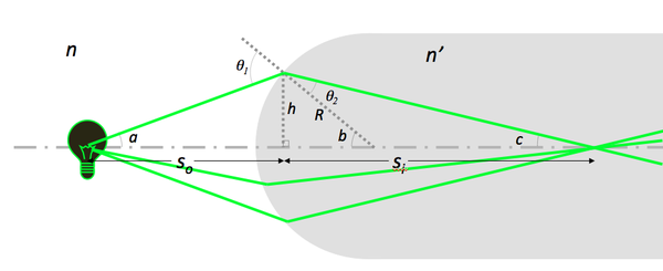
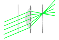
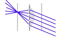
- Under Gaussian optics conditions, Snell's law predicts the image position of an object formed by a lens:
- $ n\ \sin \theta_1 = n'\ \sin \theta_2 $
- $ \sin \theta_1 \approx \sin a + \sin b \approx {h \over S_o} + {h \over R} $
- $ \sin \theta_2 \approx \sin b - \sin c \approx {h \over R} - {h \over S_i} $
- $ {n \over S_o} + {n' \over S_i} = {(n'\ - n)\over R} $
Note that
- $ S_i $ does not depend on the angle $ a $.
- Light coming from a point on the filament passes through a point after refraction.
| a) $ S_o > {n\ R \over (n'\ - n)}\ \Rightarrow S_i > 0 $ , | b) $ S_o = {n\ R \over (n'\ - n)}\ \Rightarrow S_i \to + \infty $, | c) $ S_o < {n\ R \over (n'\ - n)}\ \Rightarrow S_i < 0 $ |
Lenses
Lens maker formula
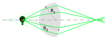
A simple lens consists of two spherical interfaces of radii of curvature $ R_1 $ and $ R_2 $. Its focal length $ f $ is given by the lens maker formula:
- $ {1 \over S_o} + {1 \over S_i} = {1 \over f} = {(n'\ - n) \over n} \left ( {1 \over R_1} - {1 \over R_2} \right ) $
| Bi-convex lens | Plano-concave lens |
|---|---|
- In your ray-tracing studies, you'll use the image from the first refraction as the object for the second.
- Note the sign convention for the second surface: $ R_2 $ < 0 for a convex lens, and $ (n'\ - n) $ has opposite sign.
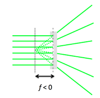
- Concave lens: the lens maker formula holds for $ R_1 $ < 0.
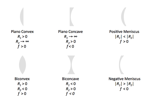
Types of spherical lenses
To minimize spherical aberrations, the general rule is to position the curved side of the lens toward the collimated/straight light.
Ray tracing
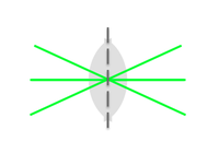
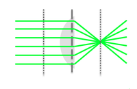
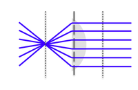
Principles
Objects and images
Imaging with a lens
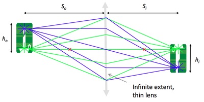
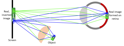
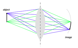
Imagine the object is made up of many point sources. After refraction, all the rays from a single point in the object plane reach the same location in the image plane. This forms a real, inverted image.
Magnification
By similar triangles, magnification is equal to the ratio of distances to the lens:
$ M = {h_i \over h_o} = {S_i \over S_o} $
You will verify this equation through the Optics Bootcamp exercise of the first problem set.
A 4 f or compound microscope
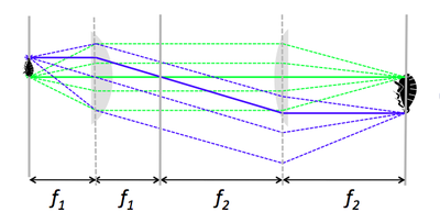
- A 4 f microscope is made of two positive lenses, placed at the sum of their focal lengths apart.
- An object (sample) placed in the focal plane of the first lens gets magnified by the ratio of focal lengths, $ f_2 / f_1 $.
- It is left as an exercise to the reader to show that the two lenses do not need to be separated by $ f_1 + f_2 $. Hint: Make clever use of Rule 3.
The eye as a lens
| |
| |
| |
You can observe a virtual image.
|
Aperture and field stops
Aperture stop and image brightness
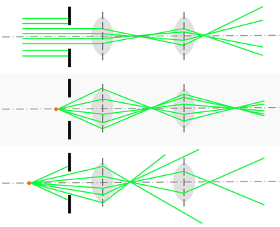
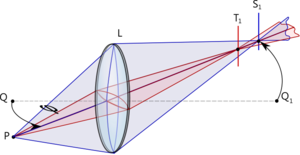
- The aperture stop of an imaging system is the optical element that limits angle of rays passing through the system from a source on the optical axis.
- For an off-axis object, the chief ray is the ray that passes through the center of the aperture stop, and the marginal rays are those that pass through the edge of the aperture stop.
- The size of the aperture stop determines the light-gathering capability of an instrument, and thus the brightness (or irradiance in W/m2) of its images.
- Typically in a microscope, as opposed to a photographic camera, the aperture stop will be the objective lens rather than an aperture before the lens as shown here.
Numerical aperture
- The numerical aperture (NA) is another important measure that characterizes the light-gathering power of a lens.
- $ NA = n\ \sin \alpha $
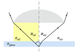
- where $ n $ is the index of refraction of the medium between the object (sample) and the lens and $ \alpha $ is the half-angle defined by the limiting ray. The limiting ray is is the last ray not blocked by any aperture.
- An oil-immersion objective lens will thus have a greater NA than an air-immersion objective lens ($ n_{oil} > n_{air} $ and $ \alpha_{oil} > \alpha_{air} $; indeed, little refraction occurs at the glass-oil interface where $ n_{oil} \approx n_{glass} $). Notice the much smaller angle of the limiting ray (drawn, but not labeled, near the point of origin in the diagram) in the case of air-immersion lens.
Field stop and field of view
- The field stop is the optical element that blocks off-axis rays passing through the center of the aperture stop (i.e. the chief rays).
- The field stop determines how much of the object can be viewed, in other words: the field of view of the imaging system.
- In your 20.309 microscope, the field stop will be the CCD camera.
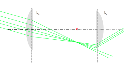
- A finite-size lens can also act as a field stop. Below is an illustration of this vignetting effect: a cone of light rays from an off-axis object is not transmitted in its entirety, but rather partially cut off by the field stop of the system, L2's rim.
Optical aberrations
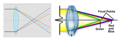
In practice, as the polychromatic nature of white light is taken into consideration, and as imaging conditions depart from the Gaussian optics approximations that have framed our representations so far, optical aberrations get introduced into images formed by lenses. Aberrations fall into two main categories: aberrations caused by wavelength variations (chromatic), and aberrations caused by the lens's spherical construction (these are known as Seidel's aberrations: spherical, coma, astigmatism, field curvature, and distortion) [1].
Chromatic aberrations
- Axial chromatic aberration is due to dispersion by the lens's medium whose index of refraction effectively varies with the light wavelength: the blue part of the spectrum is refracted to a greater extent than the red portion.
- Lateral chromatic aberration manifests itself by a difference in magnification of blue vs. red images of white-light-illuminated objects, which causes color ringing at the outer regions of the field of view.
- An achromatic doublet combination (the association of a converging lens with a weaker diverging lens) can correct chromatic aberrations for certain wavelengths.
Spherical aberrations
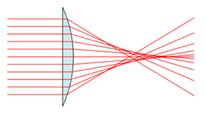
- Peripheral rays and axial rays have different focal points, because the former are actually refracted to a greater degree than the latter.
- Spherical aberrations arise from the higher-than-first-order terms in the sin θ and cos θ expansions that become non-negligible as the incident light angle θ increases.
- Spherical aberration causes the image to appear hazy or blurred and slightly out of focus.
- This effect significantly degrades the resolution of the lens because it affects the coincident imaging of points on and off the optical axis.
Coma and astigmatism
- Comatic aberration results in off-axis point objects appearing asymmetrical and taking a comet-like shape. Comatic aberration is most commonly encountered when a microscope is out of alignment.
- Astigmatism engender ellipses or blurred lines as images of speciment points. Depending on the angle of the off-axis rays entering the lens, the line image may be oriented either tangentially or radially.
Petzval distortion or field curvature
- Instead of generating image points of a flat object onto a flat screen as we have idealized so far in ray tracing estimations, a simple lens focuses these image points onto a spherical surface, shaped as a curved bowl whose curvature, the Petzval curvature, is the reciprocal of the lens radius.

