Difference between revisions of "20.109(S21):M3D4"
Noreen Lyell (Talk | contribs) (→Protocols) |
Becky Meyer (Talk | contribs) (→Part 3: Design primers for site-directed mutagenesis) |
||
| (48 intermediate revisions by one user not shown) | |||
| Line 5: | Line 5: | ||
==Introduction== | ==Introduction== | ||
| − | + | Congratulations on reaching your final (virtual) laboratory day in 20.109! To complete your experience and training, the goal for today to synthesize the data that you collected and analyzed throughout this module and refine the approach such that an improved hypothesis can be tested. By pulling all of this information together you will, hopefully, be able to use the data generated by previous 109ers to make more informed mutations that alter affinity and / or cooperativity in IPC. This module highlights the basis of scientific research as an iterative process that consists of four stages: designing experiments, collecting data, analyzing results, and refining the approach. This rigorous cycle is how we ensure our results are accurate and reproducible! | |
| − | + | When considering results that will be used to refine a research approach, it is important to recognize that not all data are created equal. For several reasons, including technical error and reagent failure, it often happens that an experiment does not work as expected. By including controls, researchers are able to identify these issues and rectify them in follow-up experiments. In addition to using controls that validate the results, researchers use replicates and repeat experiments to ensure the data are robust. All of these internal checks allow researchers to be confident about the results they report. | |
| − | + | Today you will critically think about the data that you analyzed in this module and rationally design an IPC with altered affinity / cooperativity given what you learned in your research. Though considering the results of the current Variant IPC is important in your goal for today, it is just as important to decide which results are relevant or valid to your design strategy. | |
| + | |||
| + | Though you will not have the opportunity to test your Variant IPC this semester, you are more than welcome to complete the calcium titration experiment with your protein as soon as you are invited back to campus! We will be more than happy to host you in the laboratory for some actual benchwork!! | ||
==Protocols== | ==Protocols== | ||
| − | ===Part 2: | + | ===Part 1: Review site-directed mutagenesis=== |
| + | |||
| + | Site-directed mutagenesis (SDM) refers to the a method used to incorporate specific and targeted sequence changes, or mutations, into double-stranded plasmid DNA. There are several experimental questions that can be answered by incorporating specific mutations, for example: | ||
| + | *How do amino acid substitutions alter protein / enzyme activity? | ||
| + | *How do basepair changes alter binding activity / partners at promoter sequences? | ||
| + | |||
| + | To perform SDM, custom designed oligonucleotides, or primers, are used to incorporate mutations into double-stranded DNA plasmid as a specific location in the sequence. One approach is to use primers that align to the sequence in the plasmid in a back-to-back orientation. As shown in top left of the schematic below, the primers (forward primer = black arrow and reverse primer = red arrow) anneal to the plasmid such that the 5' ends of the primers anneal to the DNA in a back-to-back orientation. In Step #1 of the schematic, the forward primer is used to replicate the top strand (outside circle of the plasmid) and the reverse primer is used to replicate the bottom strand (inside circle of the plasmid). The resulting single-stranded products (extension from each primer generates a single-stranded product) are able to anneal due to sequence homology, as shown in the first quadrant of the zoom-in for Step #2. In Step #2A the 5' ends of the linear, single-stranded amplification products are phosphorylated to prepare for ligation (Step #2B). Remember that a 5' phosphate is required for 3' OH nucleophilic attack, this results in circular plasmids. | ||
| + | |||
| + | Thus far in this description of SDM, one very important detail has not been mentioned. How specifically are the mutations coded in the primers incorporated into the plasmid sequence? In the top left of the schematic, the forward primer contains a hash mark that represents the desired mutation. The single-stranded product that results from extension from this primer will contain the desired mutation and therefore be incorporated into the products generated in Step #1. Lastly, in Step #2C the plasmid template that contains the unmutated parental sequence is destroyed so that only the plasmids with the desired mutation are present at the end of the procedure. | ||
| + | |||
| + | [[Image:Sp16 M1D3 SDM schematic.png|thumb|center|650px|'''Schematic of NEB Q5 Site Directed Mutagenesis procedure.''' Image modified from Q5 Site-Directed Mutagenesis Kit Manual published by NEB.]] | ||
| + | |||
| + | ===Part 2: Identify amino acid substitution target for new IPC design=== | ||
| + | Your first task for today is to review the data analysis you completed on M3D3 and decide which Variant IPC data you will consider when designing your Variant IPC. After you choose which amino acid you think is the best target for altering affinity / cooperativity, consider what amino acid you want to include instead. | ||
| + | |||
| + | In Part 3, you will generate the primers that can be used to incorporate a specific amino acid substitution to create your Variant IPC! | ||
| + | |||
| + | <font color = #4a9152 >'''In your laboratory notebook,'''</font color> complete the following: | ||
| + | *What amino acid will you target using SDM? At what position is this amino acid located in the protein sequence? What amino acid will be incorporated in its place? | ||
| + | *Provide the rational for your design choice. | ||
| + | **Why do you think the target amino acid you selected will alter affinity / cooperativity? | ||
| + | **How do you think the amino acid substitution will alter affinity / cooperativity? | ||
| + | |||
| + | ===Part 3: Design primers for site-directed mutagenesis=== | ||
| + | |||
| + | It is not experimentally efficient, or entirely plausible, to pick out and modify a single amino acid residue in inverse pericam post-translationally. Instead researchers genetically encode for amino acid substitutions by incorporating mutations in the DNA sequence. This is accomplished by making changes to the basepairs of a gene of interest that was cloned into a plasmid. Then the plasmid with the mutated gene is amplified using bacterial cells. | ||
| + | |||
| + | [[Image:Sp16 M1D2 primer design schematic.png|thumb|right|300px| '''Schematic for mutating gene sequences in plasmids using SDM technique.''' Image modified from Q5 Site-Directed Mutagenesis Kit Manual published by NEB.]]To incorporate a mutation at a specific location in the DNA sequence, synthetic primers can be used in a technique referred to site-direction mutagenesis (see figure on the right). Primer design for site-directed mutagenesis, or SDM, is quite straightforward: the forward primer introduces a mutation into the coding strand. Both non-mutagenic and mutagenic amplification require cycles of DNA melting, annealing, and extension. | ||
| + | |||
| + | Primers used in SDM must meet several design criteria to ensure specificity and efficiency. Consider the following design guidelines for mutagenesis primers: | ||
| + | |||
| + | *Desired mutation (1-2 bp) must be present in the middle of the forward primer. | ||
| + | *Forward and reverse primers should 'face' away from the mutation and be 'back-to-back' when annealed to the template. | ||
| + | *Primers should be 25-45 bp long. | ||
| + | *G/C content of > 40% is desired. | ||
| + | *Both primers should terminate in at least one G or C base. | ||
| + | *Melting temperature should exceed 78°C, according to: | ||
| + | **T<sub>m</sub> = 81.5 + 0.41 (%GC) – 675/N - %mismatch | ||
| + | **where N is primer length and the two percentages should be integers | ||
| + | |||
| + | To demonstrate primer design, the illustration below uses S101L, which is an uninteresting mutation but a helpful example: | ||
| + | |||
| + | Residue 101 of calmodulin is serine, encoded by the AGC codon. This is residue 379 with respect to the entire inverse pericam construct, | ||
| + | and we can find it and some flanking code in the DNA sequence from Part 2: | ||
| + | |||
| + | <font face="courier"> | ||
| + | <small> | ||
| − | + | 361 (5') GAG GAA ATC CGA GAA GCA TTC CGT GTT TTT GAC AAG GAT GGG AAC GGC TAC ATC AGC GCT (3') | |
| − | + | 381 (5') GCT CAG TTA CGT CAC GTC ATG ACA AAC CTC GGG GAG AAG TTA ACA GAT GAA GAA GTT GAT (3') | |
| − | < | + | </small> |
| − | + | </font> | |
| − | + | ||
| − | = | + | To change from serine to leucine, one might choose TTA, TTG, or CTN (wherer N = T, A, G, or C). Because CTC requires only two mutations (rather than three as for the other options), we choose this codon. |
| − | [ | + | Now we must keep >10 bp of sequence on each side in a way that meets all our requirements. To quickly find G/C content and see secondary structures, look at the [https://www.idtdna.com/pages/tools/oligoanalyzer IDT website]. (Note that the T<sub>m</sub> listed at this site is '''''not''''' one that is relevant for mutagenesis.) |
| − | + | Ultimately, your forward primer might look like the following, which has a T<sub>m</sub> of almost 81°C, and a G/C content of ~58%. | |
| − | + | ||
| − | + | ||
| − | + | ||
| − | + | ||
| − | + | ||
| − | + | ||
| − | + | ||
| − | + | ||
| − | = | + | <font face="courier"> |
| + | 5’ GG AAC GGC TAC ATC CTC GCT GCT CAG TTA CGT CAC G 3' | ||
| + | </font><br> | ||
| − | + | The reverse primer is the inverse complement of a sequence just preceding the forward primer in the IPC gene. The forward and reverse primers are set up back-to-back. | |
| − | + | ||
| − | + | ||
| − | = | + | Luckily, online tools are available to assist with SDM primer design. Today you will use NEBaseChanger (provided by NEB) to design your mutagenic primers. |
| + | #Go to the [http://nebasechanger.neb.com/ NEBaseChanger] site and click 'Please enter a new sequence to begin.' | ||
| + | #*A new window will open. | ||
| + | #Copy and paste the WT IPC sequence. | ||
| + | #*This sequence should be saved in SnapGene from the M3D1 exercise. Alternatively, you can copy the sequence from the word document attached to the M3D1 wiki page. | ||
| + | #Confirm that the 'Substitution' option is selected. | ||
| + | #Highlight the basepairs you want to mutate using by scrolling through the sequence, or you can search the sequence by typing the basepairs into the 'Find' box. | ||
| + | #Type the new DNA sequence (the basepair(s) you want your forward mutagenic primer to incorporate into the IPC sequence) in the 'Desired Sequence' box. | ||
| + | #*Under the Result header, a diagram showing where your primers will anneal is provided. | ||
| + | #*Under the Required Primers header, the sequences for your forward primer and reverse primer are shown with the characteristics for each. | ||
| + | #<font color = #4a9152 >'''In your laboratory notebook,'''</font color> complete the following: | ||
| + | #*Include a screen capture of the information provided in the Result and Required Primers sections. | ||
| + | #*Use the guidelines provided above to examine the mutagenesis primers designed by NEBaseChanger. Do the primers meet the design criteria? | ||
| + | ===Part 4: Start Mini-report assignment=== | ||
| − | + | The final writing assignment in Module 3 is the Mini-report. In this assignment you will provide a description of the data analysis you completed to study the effects of mutating residues in IPC. To help you organize your thoughts and to ensure you ready to prepare this assignment in the next laboratory session, '''work with your laboratory partner(s)''' to draft an outline of your Mini-report. Before you prepare your outline, review the [[20.109(S21):Mini-report |Mini-report assignment page]] for guidance! | |
| − | + | ||
| − | + | ||
| − | * | + | <font color = #4a9152 >'''In your laboratory notebook,'''</font color> complete the following: |
| + | *List the key topics that will be addressed / explained in the Background and Approach section. | ||
| + | *List the figures that will be included and provide a brief description of how the data will be presented in the figures. | ||
| + | **Not all of the figures that were generated are required for this assignment! Carefully consider which data are useful / important in the rational design of your new Variant IPC. | ||
| + | *Include a statement concerning the interpretation of the data represented in each figure. | ||
==Navigation links== | ==Navigation links== | ||
| − | + | Previous day: [[20.109(S21):M3D3 |Evaluate effect of mutations on IPC variants ]] <br> | |
| − | Previous day: | + | |
Latest revision as of 18:30, 11 May 2021
Contents
Introduction
Congratulations on reaching your final (virtual) laboratory day in 20.109! To complete your experience and training, the goal for today to synthesize the data that you collected and analyzed throughout this module and refine the approach such that an improved hypothesis can be tested. By pulling all of this information together you will, hopefully, be able to use the data generated by previous 109ers to make more informed mutations that alter affinity and / or cooperativity in IPC. This module highlights the basis of scientific research as an iterative process that consists of four stages: designing experiments, collecting data, analyzing results, and refining the approach. This rigorous cycle is how we ensure our results are accurate and reproducible!
When considering results that will be used to refine a research approach, it is important to recognize that not all data are created equal. For several reasons, including technical error and reagent failure, it often happens that an experiment does not work as expected. By including controls, researchers are able to identify these issues and rectify them in follow-up experiments. In addition to using controls that validate the results, researchers use replicates and repeat experiments to ensure the data are robust. All of these internal checks allow researchers to be confident about the results they report.
Today you will critically think about the data that you analyzed in this module and rationally design an IPC with altered affinity / cooperativity given what you learned in your research. Though considering the results of the current Variant IPC is important in your goal for today, it is just as important to decide which results are relevant or valid to your design strategy.
Though you will not have the opportunity to test your Variant IPC this semester, you are more than welcome to complete the calcium titration experiment with your protein as soon as you are invited back to campus! We will be more than happy to host you in the laboratory for some actual benchwork!!
Protocols
Part 1: Review site-directed mutagenesis
Site-directed mutagenesis (SDM) refers to the a method used to incorporate specific and targeted sequence changes, or mutations, into double-stranded plasmid DNA. There are several experimental questions that can be answered by incorporating specific mutations, for example:
- How do amino acid substitutions alter protein / enzyme activity?
- How do basepair changes alter binding activity / partners at promoter sequences?
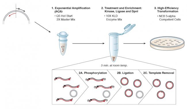
To perform SDM, custom designed oligonucleotides, or primers, are used to incorporate mutations into double-stranded DNA plasmid as a specific location in the sequence. One approach is to use primers that align to the sequence in the plasmid in a back-to-back orientation. As shown in top left of the schematic below, the primers (forward primer = black arrow and reverse primer = red arrow) anneal to the plasmid such that the 5' ends of the primers anneal to the DNA in a back-to-back orientation. In Step #1 of the schematic, the forward primer is used to replicate the top strand (outside circle of the plasmid) and the reverse primer is used to replicate the bottom strand (inside circle of the plasmid). The resulting single-stranded products (extension from each primer generates a single-stranded product) are able to anneal due to sequence homology, as shown in the first quadrant of the zoom-in for Step #2. In Step #2A the 5' ends of the linear, single-stranded amplification products are phosphorylated to prepare for ligation (Step #2B). Remember that a 5' phosphate is required for 3' OH nucleophilic attack, this results in circular plasmids.
Thus far in this description of SDM, one very important detail has not been mentioned. How specifically are the mutations coded in the primers incorporated into the plasmid sequence? In the top left of the schematic, the forward primer contains a hash mark that represents the desired mutation. The single-stranded product that results from extension from this primer will contain the desired mutation and therefore be incorporated into the products generated in Step #1. Lastly, in Step #2C the plasmid template that contains the unmutated parental sequence is destroyed so that only the plasmids with the desired mutation are present at the end of the procedure.
Part 2: Identify amino acid substitution target for new IPC design
Your first task for today is to review the data analysis you completed on M3D3 and decide which Variant IPC data you will consider when designing your Variant IPC. After you choose which amino acid you think is the best target for altering affinity / cooperativity, consider what amino acid you want to include instead.
In Part 3, you will generate the primers that can be used to incorporate a specific amino acid substitution to create your Variant IPC!
In your laboratory notebook, complete the following:
- What amino acid will you target using SDM? At what position is this amino acid located in the protein sequence? What amino acid will be incorporated in its place?
- Provide the rational for your design choice.
- Why do you think the target amino acid you selected will alter affinity / cooperativity?
- How do you think the amino acid substitution will alter affinity / cooperativity?
Part 3: Design primers for site-directed mutagenesis
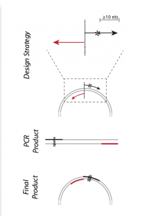
It is not experimentally efficient, or entirely plausible, to pick out and modify a single amino acid residue in inverse pericam post-translationally. Instead researchers genetically encode for amino acid substitutions by incorporating mutations in the DNA sequence. This is accomplished by making changes to the basepairs of a gene of interest that was cloned into a plasmid. Then the plasmid with the mutated gene is amplified using bacterial cells.
Primers used in SDM must meet several design criteria to ensure specificity and efficiency. Consider the following design guidelines for mutagenesis primers:
- Desired mutation (1-2 bp) must be present in the middle of the forward primer.
- Forward and reverse primers should 'face' away from the mutation and be 'back-to-back' when annealed to the template.
- Primers should be 25-45 bp long.
- G/C content of > 40% is desired.
- Both primers should terminate in at least one G or C base.
- Melting temperature should exceed 78°C, according to:
- Tm = 81.5 + 0.41 (%GC) – 675/N - %mismatch
- where N is primer length and the two percentages should be integers
To demonstrate primer design, the illustration below uses S101L, which is an uninteresting mutation but a helpful example:
Residue 101 of calmodulin is serine, encoded by the AGC codon. This is residue 379 with respect to the entire inverse pericam construct, and we can find it and some flanking code in the DNA sequence from Part 2:
361 (5') GAG GAA ATC CGA GAA GCA TTC CGT GTT TTT GAC AAG GAT GGG AAC GGC TAC ATC AGC GCT (3')
381 (5') GCT CAG TTA CGT CAC GTC ATG ACA AAC CTC GGG GAG AAG TTA ACA GAT GAA GAA GTT GAT (3')
To change from serine to leucine, one might choose TTA, TTG, or CTN (wherer N = T, A, G, or C). Because CTC requires only two mutations (rather than three as for the other options), we choose this codon.
Now we must keep >10 bp of sequence on each side in a way that meets all our requirements. To quickly find G/C content and see secondary structures, look at the IDT website. (Note that the Tm listed at this site is not one that is relevant for mutagenesis.)
Ultimately, your forward primer might look like the following, which has a Tm of almost 81°C, and a G/C content of ~58%.
5’ GG AAC GGC TAC ATC CTC GCT GCT CAG TTA CGT CAC G 3'
The reverse primer is the inverse complement of a sequence just preceding the forward primer in the IPC gene. The forward and reverse primers are set up back-to-back.
Luckily, online tools are available to assist with SDM primer design. Today you will use NEBaseChanger (provided by NEB) to design your mutagenic primers.
- Go to the NEBaseChanger site and click 'Please enter a new sequence to begin.'
- A new window will open.
- Copy and paste the WT IPC sequence.
- This sequence should be saved in SnapGene from the M3D1 exercise. Alternatively, you can copy the sequence from the word document attached to the M3D1 wiki page.
- Confirm that the 'Substitution' option is selected.
- Highlight the basepairs you want to mutate using by scrolling through the sequence, or you can search the sequence by typing the basepairs into the 'Find' box.
- Type the new DNA sequence (the basepair(s) you want your forward mutagenic primer to incorporate into the IPC sequence) in the 'Desired Sequence' box.
- Under the Result header, a diagram showing where your primers will anneal is provided.
- Under the Required Primers header, the sequences for your forward primer and reverse primer are shown with the characteristics for each.
- In your laboratory notebook, complete the following:
- Include a screen capture of the information provided in the Result and Required Primers sections.
- Use the guidelines provided above to examine the mutagenesis primers designed by NEBaseChanger. Do the primers meet the design criteria?
Part 4: Start Mini-report assignment
The final writing assignment in Module 3 is the Mini-report. In this assignment you will provide a description of the data analysis you completed to study the effects of mutating residues in IPC. To help you organize your thoughts and to ensure you ready to prepare this assignment in the next laboratory session, work with your laboratory partner(s) to draft an outline of your Mini-report. Before you prepare your outline, review the Mini-report assignment page for guidance!
In your laboratory notebook, complete the following:
- List the key topics that will be addressed / explained in the Background and Approach section.
- List the figures that will be included and provide a brief description of how the data will be presented in the figures.
- Not all of the figures that were generated are required for this assignment! Carefully consider which data are useful / important in the rational design of your new Variant IPC.
- Include a statement concerning the interpretation of the data represented in each figure.


