Difference between revisions of "20.109:Module 2"
(→Module 2) |
|||
| Line 9: | Line 9: | ||
'''TA:''' [[Yoon Sung Nam]] | '''TA:''' [[Yoon Sung Nam]] | ||
| − | In this | + | Once proteins are synthesized, their localization and conformation are critical to their function, but without “X-ray vision” to peer into a cell, these can be hard to detect. Fluorescence is one of the best tools in our toolkit for measuring proteins in living cells. By tagging a protein of interest with a fluorescent one, cellular functions can be probed and observed unobtrusively. In this experiment we will follow not a protein of interest but a cellular signal, namely calcium fluctuations. We will transfect a genetically-encoded calcium sensor into mouse embryonic stem cells. In the presence of calcium, the fluorescent protein will fold and the cells should appear green. Changes in fluorescence will allow us to quantitatively measure how chemical and physical perturbations affect calcium signaling in these cells. |
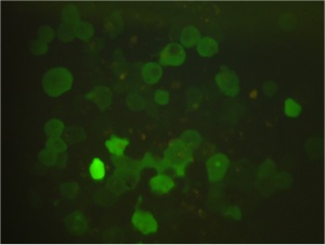
[[Image:Macintosh HD-Users-nkuldell-Desktop-SgnlMeasrmnt coverart S07.jpg|thumb|300px|center|Mouse embryonic stem cells expressing genetically-encoded Ca2+ sensor<br> image from N. Kuldell]] | [[Image:Macintosh HD-Users-nkuldell-Desktop-SgnlMeasrmnt coverart S07.jpg|thumb|300px|center|Mouse embryonic stem cells expressing genetically-encoded Ca2+ sensor<br> image from N. Kuldell]] | ||
Revision as of 11:11, 27 November 2006
Module 2
Instructors: Alan Jasanoff and Natalie Kuldell
TA: Yoon Sung Nam
Once proteins are synthesized, their localization and conformation are critical to their function, but without “X-ray vision” to peer into a cell, these can be hard to detect. Fluorescence is one of the best tools in our toolkit for measuring proteins in living cells. By tagging a protein of interest with a fluorescent one, cellular functions can be probed and observed unobtrusively. In this experiment we will follow not a protein of interest but a cellular signal, namely calcium fluctuations. We will transfect a genetically-encoded calcium sensor into mouse embryonic stem cells. In the presence of calcium, the fluorescent protein will fold and the cells should appear green. Changes in fluorescence will allow us to quantitatively measure how chemical and physical perturbations affect calcium signaling in these cells.
Module 2 Day 1: Start-up signal measurement
Module 2 Day 2: Measuring calcium in vitro
Module 2 Day 3: Lipofection
Module 2 Day 4: Measuring calcium in vivo
Module 2 Day 5:Calcium signaling in vivo
Module 2 Day 6: Student presentations

