20.109(S17): Laboratory Fundamentals of Biological Engineering
Introduction
In the previous laboratory session you screened SMM slides using your purified FKBP12 protein. To identify which of the small molecules you screened bind FKBP12, your slides will be evaluated using a Genepix microarray scanner. The scanner will measure the fluorescence signal emitted from each spot on the slide where a ligand was printed. Considering that neither the ligands nor FKBP12 protein is fluorescent, from where does this signal arise?
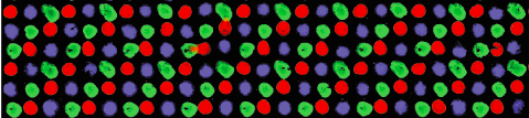
The image to the right is from a pilot experiment completed in preparation for this module. The green spots represent locations on the SMM slide where fluorescein was printed. Fluorescein is a fluorescent dye used for alignment purposes. A positive control was also included on each slide. You may remember that 4 x 48 spots were printed with rapamycin. Rapamycin is a macrolide compound used to suppress the immune system in organ transplant recipients. For our purposes, it is a compound known to bind FKBP12. In the image to the right, the bright red spots show where FKBP12 bound to the rapamycin at these locations. However, this still doesn't answer the question concerning the source of the red fluorescent signal. Think back to the purification procedure. A His tag was used to isolate FKBP12 from the cell lysate. This tag can also act as a target for antibody probes. The anti-His antibody that you added to your slides attached to the His-tagged FKBP12 protein that were bound to the slide (more specifically, the ligands that were linked to the slides). The anti-His antibody has two important features: it is able to bind a His tag and it carries an fluorophore. As shown in the image below, the ligand-protein-antibody complex results in a fluorophore at only sites where FKBP12 binding occurs on the slide.


