20.109(F07): Luciferase assays and RNA prep
Contents
Introduction
Facts vs Data...plus a measure of measurement
Three basic questions drive all scientific efforts: what’s here, how did it get here, and how does it work…three simple questions that have given rise to a mind-numbing number of textbooks, many of which are themselves mind-numbing. How sad. It’s too easy to open a science textbook and read statements that seem either obvious (e.g. all living things are made of cells) or unbelievably detailed (e.g. poliovirus is a positive-stranded RNA virus so its genome can serve as mRNA). Scientific ideas, even the “no duh” ones, rely on evidence that has been extensively examined and logically interpreted. That’s what makes it science. There may be other ideas and explanations for how things work but when the preponderance of evidence supports an idea, it becomes the science. The process of discovery, driven by data collection and interpretation, is such an integral part of science that it’s surprising how rarely it gets taught. It’s easy to forget that science is a human endeavor, advanced by curious and hardworking individuals and communities, all hoping to improve our understanding of the natural world.
Aristotle (~300 BC) observed nature directly and since then science has presumed that we can learn about the world by collecting evidence. Gathering data has become so integral to science that it seems an obvious part. However when Aristotle started looking at the world, he concluded that
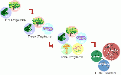
- all organisms were either animals or plants
- plants were less complex than animals and so ranked “lower” on some life ladder
- living things spontaneously arise from non-living things
Read that last point again. Spontaneous generation seemed a sensible idea to wise old Aristotle based on what was observable. And amazingly this completely wrong theory was in place for nearly 2000 years. Rudolph Virchow didn’t state that “where a cell exists, there must have been a preexisting cell” until 1858…not even 200 years ago!
Coupled with data collection is instrumentation and technology. Advances in technology often lead to scientific progress. (e.g. microscopes that heralded the cell theory) and technology also depends on improved scientific descriptions (e.g. the wave and particle theories of light leading to the development of new microscopes). Instruments serve as extensions of our five senses to gather data. As such, new instruments can be like new ways of testing and seeing the world.
Scientific work generates evidence-based, internally consistent, and well-tested explanations. Gathering data is a big part of the fun and a big part of what you’ll do over the next few lab sessions. In today’s lab we will collect direct evidence about the ability of the siRNA you’ve designed to reduce the expression of the Renilla luciferase gene. Next time you will examine other genes whose expression pattern may have changed as a result of your siRNA. Together these lines of evidence, collected with instruments that can “see” what our eyes can’t, will help you explain what the cells have done in response to your experiment.
Protocols
Part 1: Preparing cell lysates
RNA is strikingly different from DNA in its stability. Consequently it is more difficult to work with RNA in the lab. It is not the techniques themselves that are difficult; indeed, many of the manipulations will seem identical to those used for DNA. However, RNA is rapidly and easily degraded by RNases that exist everywhere. There are several rules for working with RNA. They will improve your chances of success. Please follow them all.
- Use warm water on a paper towel to wash lab equipment, like microfuges, before you begin your experiment. Then wipe them down with “RNase-away” solution.
- Wear gloves when you are touching anything that will touch your RNA.
- Change your gloves often.
- Before you begin your experiment clean your work area, removing all clutter. Wipe down the benchtop with warm water then “RNase-away,” and then lay down a fresh piece of benchpaper.
- Use RNA-dedicated solutions and if possible RNA-dedicated pipetmen.
- Start a new box of pipet tips and label their lid “RNA ONLY.”
Qiagen sells a kit for isolating RNA and we will be using their protocol and reagents.
- Retrieve your cells from the TC facility (be sure to get both the transfection plates and the 6-well dish that you seeded during "cell culture for fun") and take them to the main teaching lab.
- Take digital photographs of the cells in wells transfected with plasmid and siRNAs. Email these photographs to you and your lab partner or post them to your userpage. While you are waiting for the camera, you can examine the 6-well dish that has been growing for one week and write any observations about the wells in your lab notebook.
- Aspirate the media from the cells of your six-well dishes and wash 2X with 2 ml PBS.
- Lyse the cells in each well by adding 500 ul PLB, a reagent sold by Promega that will lyse the cells into a buffer compatible with the luciferase assay reagents. Incubate on the orbital shaker in the main teaching lab for 15 minutes at 150 rpm.
- Move the lysates to RNase-free eppendorf tubes with clean pipet tips. Remove 100 ul of each to use in the luciferase assays.
Part 2: Luciferase assays
These assays should be performed as you did last time. Refer to that protocol for any particulars of the assay that you may have forgotten.
Part 3: Isolation of total RNA
- Add 400 ul of Buffer RTL with BME to the two experimental samples you will study by microarray. This should be done in the fume hood since the reagent contains β-mercaptoethanol which smells awful.
- Collect two Qiashredder columns from one of the teaching faculty. The lysates must be passed through these columns to remove particulate matter. Load the top of each Qiashredder column with the cell lysates. The remainder of today’s experiment can be performed in the main teaching lab, but remember to wear gloves throughout to protect your RNA samples from RNases on your hands.
- Microfuge the Qiashredder columns for 2 minutes. Move the flow-through into two properly-labeled eppendorf tubes.
- Add 1 volume (approximately 600 ul) of 70% ethanol to each of the cleared lysates. Invert 3 or 4 times to mix the contents. Do not vortex. A precipitate may form at this stage but it will not effect your RNA isolation.
- Collect two Rneasy minicolumns from the teaching faculty. Apply 700 ul of each sample to the columns and microfuge for 15 seconds. Discard the flow-through. Keep the collection tube.
- Apply any remaining sample to the columns. Microfuge and discard the flow-through as before.
- Add 700 ul Buffer RW1 to each column. Microfuge for 15 seconds. Discard both the flow-through and the collection tube.
- Transfer each column to a fresh 2 ml collection tube and add 500 ul Buffer RPE to each column. Microfuge for 15 seconds and discard the flow-through.
- Add another 500 ul Buffer RPE to each column and microfuge for 2 minutes. Discard the flow-through and the collection tube.
- Transfer each column to a fresh collection tube and microfuge for 1 minute. This step is important since it removes any residual ethanol from the membrane.
- Trim the cap off two new 1.5 ml eppendorf tubes (save the caps!) and label the sides of the tubes with your team color, the date and a name for the sample. Transfer the columns into the trimmed eppendorf tubes and elute the RNA from the columns by adding 50 ul of RNase-free water to each. Microfuge for 1 minute then cap and store the samples on ice.
Part 4: Measure RNA concentration
- Measure the concentration of your RNA sample by adding 5 μl to 495 μl sterile water. The water does not have to be RNase-free since the RNA can be degraded and still give legitimate readings in the spectrophotometer. Make your dilutions in an eppendorf tube and use your P1000 to transfer the dilution to a quartz cuvette. Measure the absorbance at 260 nm. Water in one of the optically paired cuvettes should be used to blank the spectrophotometer, but if another group has done this already, it does not have to be repeated.
- A few things to be aware of when using quartz cuvettes:
- They are very expensive.
- The lab has only one set.
- When you are done using the cuvette, you should carefully clean it by shaking out the contents into the sink and rinsing it once with 70% EtOH, then two times with water. Quartz cuvettes get most of their chips and cracks when someone is shaking out the contents since it is so easy for the cuvette to slip from wet fingers or be hit against the sink. Don’t let this happen to you.
- A few things to be aware of when using quartz cuvettes:
- To determine the concentration of RNA in your sample, use the fact that 40 μg/ml of RNA will give a reading of 1 A260.
| RNA Sample | A260 | Conc of dilute RNA | Conc of undiluted RNA |
|---|---|---|---|
| control sample | |||
| variable sample |
DONE!
For next time
- Post your luciferase data to the talk page associated with today's lab. This will be important so others in the lab can consider your results in addition to their own and so the team in the other lab who used the same siRNA can consider the reproducibility of their work. This analysis will be included in your written report for this module.
- Analyze your luciferase data as you did last time, namely with a bar graph comparing each sample's ALU. You might want to average the duplicates if they are close or show individual replicates if they are not. Be sure to explicitly state what can you conclude about the efficacy of each siRNA. This figure will become part of your written report for this module.
- Please download the midsemester evaluation form found here. Complete the questionnaire and then print it out without including your name to turn in next time. If there is something you'd like to see done differently for the rest of the course, this is your chance to lobby for that change. Similarly, if there is something you think the class has to keep doing, let us know that too.
- Read the following article for a class discussion next time.
- The news story by Erika Check called "RNA interference: hitting the on switch" published in Nature 2007 vol. 448 pp. 855-8
- The original paper describing some work labeled "RNA activation" by Long-Cheng Li, et al. The article is titled: Small dsRNAs induce transcriptional activation in human cells and was published in PNAS 2006 vol. 103 pp. 17337-42.
- Questions to guide your reading and some background information can be found on the page associated with the next lab. You do NOT have to turn in the answers to these questions but you will be asked to guide the class discussion around some of these topics so you must come to lab familiar with the content of the articles and ready to discuss the material.
Reagents list
- PLB
- Promega reagent (“Passive Lysis Buffer”)
- Buffer RTL/BME
- Qiagen reagent with BME
- Buffer RW1
- Qiagen reagent
- Buffer RPE
- Qiagen reagent with ethanol


