Difference between revisions of "Spring 2012:Leanna Morinishi Lit Search"
From Course Wiki
(→Light Field Microscopy) |
(→Light Field Microscopy) |
||
| Line 4: | Line 4: | ||
= Light Field Microscopy = | = Light Field Microscopy = | ||
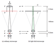
Light field imaging allows for the capture of the light field of a sample in a single photograph.<ref>[http://graphics.stanford.edu/software/LFDisplay/lfmintro.pdf A practical introduction to light field microscopy - Stanford 2010 (pdf)]</ref> This has been recently commercialized in the [http://www.lytro.com/living-pictures/1699 Lytro Camera]<ref>[http://graphics.stanford.edu/papers/lfcamera/ Light field camera]</ref> which obtains a light field using a microlens and allows for processing of the image afterwards.[[File:LightFieldMic.png|thumb|Light Field Microscope Diagram from the Stanford 2006 paper.<ref>[http://graphics.stanford.edu/papers/lfmicroscope/levoy-lfmicroscope-sig06.pdf Light field microscopy - Stanford 2006 (pdf)]</ref>]] | Light field imaging allows for the capture of the light field of a sample in a single photograph.<ref>[http://graphics.stanford.edu/software/LFDisplay/lfmintro.pdf A practical introduction to light field microscopy - Stanford 2010 (pdf)]</ref> This has been recently commercialized in the [http://www.lytro.com/living-pictures/1699 Lytro Camera]<ref>[http://graphics.stanford.edu/papers/lfcamera/ Light field camera]</ref> which obtains a light field using a microlens and allows for processing of the image afterwards.[[File:LightFieldMic.png|thumb|Light Field Microscope Diagram from the Stanford 2006 paper.<ref>[http://graphics.stanford.edu/papers/lfmicroscope/levoy-lfmicroscope-sig06.pdf Light field microscopy - Stanford 2006 (pdf)]</ref>]] | ||
| + | |||
| + | I would be thrilled to work on this project for a number of reasons: | ||
| + | * It will force me to work on image processing, which I am not good at :p | ||
| + | * It would be an awesome project | ||
| + | * It is '''feasible''' | ||
| + | ** Only one additional physical component in a traditional bright field microscope | ||
| + | ** Lots of code | ||
| + | * It is '''relevant''' | ||
| + | ** This microscope can be used to compose a 3-dimensional image of the sample | ||
| + | ** Taking 3-dimensional images quickly is a current point of interest in microscopy today | ||
| + | * It is '''novel''' | ||
| + | ** With respect to this class, this is a novel project | ||
| + | ** Otherwise, this is not a new technology <ref name="1996"/> but has been implemented into prototype microscopes in the past 7 years | ||
They also documented their setup in more detail in a technical memo.<ref>[http://graphics.stanford.edu/papers/lfmicroscope/lfmicroscope-optics.pdf Optical recipes for light field microscopes - Stanford 2006 (pdf)]</ref> | They also documented their setup in more detail in a technical memo.<ref>[http://graphics.stanford.edu/papers/lfmicroscope/lfmicroscope-optics.pdf Optical recipes for light field microscopes - Stanford 2006 (pdf)]</ref> | ||
| Line 9: | Line 22: | ||
Introduction to LFDisplay.<ref>[http://graphics.stanford.edu/software/LFDisplay/ LFDisplay: A real time system for light field microscopy]</ref> | Introduction to LFDisplay.<ref>[http://graphics.stanford.edu/software/LFDisplay/ LFDisplay: A real time system for light field microscopy]</ref> | ||
| − | Light field rendering.<ref>[http://graphics.stanford.edu/papers/light/ Light field rendering - Stanford 1996]</ref> | + | Light field rendering.<ref name="1996">[http://graphics.stanford.edu/papers/light/ Light field rendering - Stanford 1996]</ref> |
In 2009, the group published a better quality machine 4D Microscopy<ref>[http://graphics.stanford.edu/papers/lfillumination/levoy-lfillumination-jmicr09.pdf Recording and controlling the 4D light field in a microscope - Stanford 2009 (pdf)]</ref> | In 2009, the group published a better quality machine 4D Microscopy<ref>[http://graphics.stanford.edu/papers/lfillumination/levoy-lfillumination-jmicr09.pdf Recording and controlling the 4D light field in a microscope - Stanford 2009 (pdf)]</ref> | ||
Revision as of 17:23, 5 March 2012
Light Field Microscopy
Light field imaging allows for the capture of the light field of a sample in a single photograph.[1] This has been recently commercialized in the Lytro Camera[2] which obtains a light field using a microlens and allows for processing of the image afterwards.
Light Field Microscope Diagram from the Stanford 2006 paper.[3]
I would be thrilled to work on this project for a number of reasons:
- It will force me to work on image processing, which I am not good at :p
- It would be an awesome project
- It is feasible
- Only one additional physical component in a traditional bright field microscope
- Lots of code
- It is relevant
- This microscope can be used to compose a 3-dimensional image of the sample
- Taking 3-dimensional images quickly is a current point of interest in microscopy today
- It is novel
- With respect to this class, this is a novel project
- Otherwise, this is not a new technology Cite error: Invalid
<ref>tag;
name cannot be a simple integer. Use a descriptive title but has been implemented into prototype microscopes in the past 7 years
They also documented their setup in more detail in a technical memo.[4]
Introduction to LFDisplay.[5]
Light field rendering.Cite error: Invalid <ref> tag;
name cannot be a simple integer. Use a descriptive title
In 2009, the group published a better quality machine 4D Microscopy[6]
References
- ↑ A practical introduction to light field microscopy - Stanford 2010 (pdf)
- ↑ Light field camera
- ↑ Light field microscopy - Stanford 2006 (pdf)
- ↑ Optical recipes for light field microscopes - Stanford 2006 (pdf)
- ↑ LFDisplay: A real time system for light field microscopy
- ↑ Recording and controlling the 4D light field in a microscope - Stanford 2009 (pdf)