Difference between revisions of "Spring 2011:3D PSF lab:Kristin"
Kristin Kuhn (Talk | contribs) |
Kristin Kuhn (Talk | contribs) |
||
| Line 6: | Line 6: | ||
<small>3D equivalent of a rect function and its Fourier transform, the PSF. ''source: www.projectrhea.org''</small> | <small>3D equivalent of a rect function and its Fourier transform, the PSF. ''source: www.projectrhea.org''</small> | ||
| − | Theoretically, then, we should be able to ''deconvolve'' the image on our computer with this PSF, and get the "true" image with much higher resolution. This PSF | + | Theoretically, then, we should be able to ''deconvolve'' the image on our computer with this PSF, and get the "true" image with much higher resolution. This PSF was obtained by taking an z-stack image of an approximate point source. |
| − | ImageJ | + | Using ImageJ, this PSF and the some camera images of intestinal cells were deconvolved, as per [[Spring_2011:3D_PSF_lab#ImageJ_Tutorial:_Deconvolution_Lab | Emmanuel's tutorial]]. I used the Richardson-Lucy algorithm, 10 iterations. |
| Line 15: | Line 15: | ||
Left: original image. Right: deconvolved image. | Left: original image. Right: deconvolved image. | ||
| − | [[File:Psf-compare-0.png|none| | + | [[File:Psf-compare-0.png|none|900px]] |
| − | [[File:Psf-compare-1.png|none| | + | [[File:Psf-compare-1.png|none|900px]] |
| − | [[File:Psf-compare-2.png|none| | + | [[File:Psf-compare-2.png|none|900px]] |
| − | [[File:Psf-compare-3.png|none| | + | [[File:Psf-compare-3.png|none|900px]] |
Latest revision as of 03:50, 19 May 2011
Because lenses aren't infinite, any time a camera looks at an image, it is like the image is being multiplied with a rect function (in 1-D). In Fourier space, the spectrum of the image is convolved with a sinc function. This reduces your resolution, blurring sharp edges into smooth curves.
In 3D, this translates to the image being convolved with the point spread function.
3D equivalent of a rect function and its Fourier transform, the PSF. source: www.projectrhea.org
Theoretically, then, we should be able to deconvolve the image on our computer with this PSF, and get the "true" image with much higher resolution. This PSF was obtained by taking an z-stack image of an approximate point source.
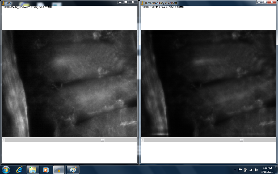
Using ImageJ, this PSF and the some camera images of intestinal cells were deconvolved, as per Emmanuel's tutorial. I used the Richardson-Lucy algorithm, 10 iterations.
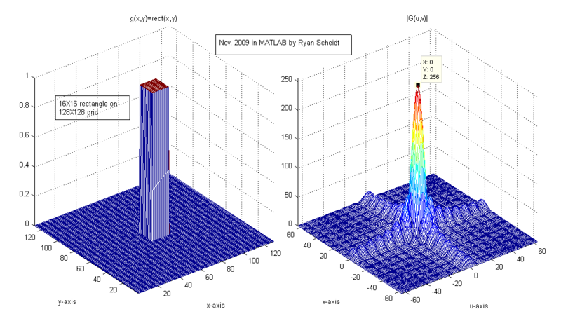
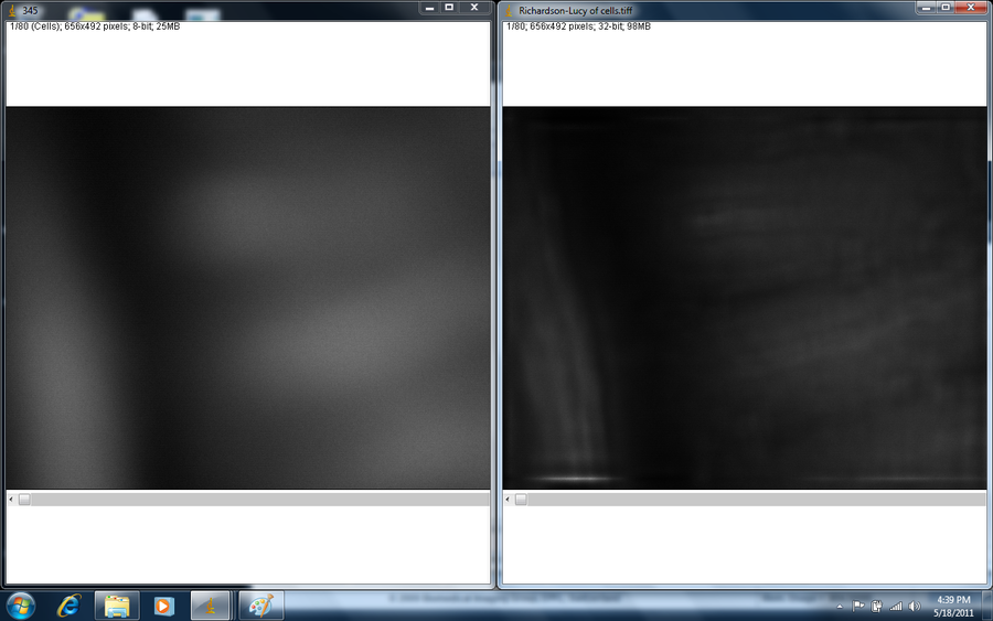
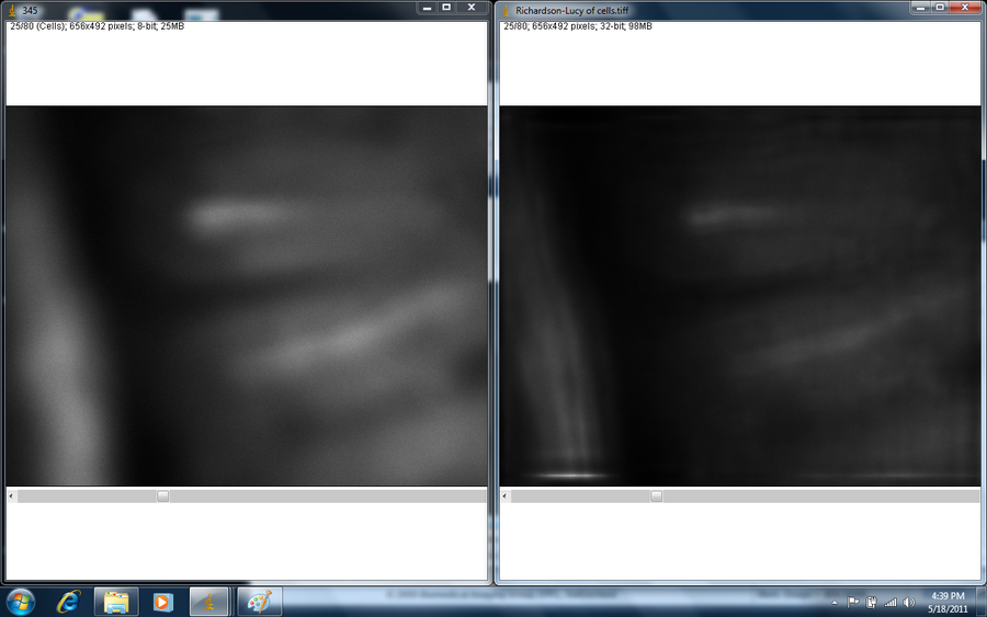
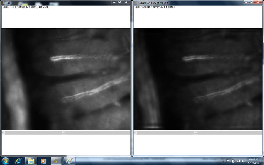
Before/after images:
Left: original image. Right: deconvolved image.
The improvement is especially noticeable in the first few frames. Even in the fourth image, you can see the increased resolution for the nodules of light in the elongated structure on the left. Note the frame of high-frequency noise around the deconvolved image.
So why don't we do this all the time? Unfortunately, it is hard to eliminate noise with this method, because once you filter an image for noise, the PSF may no longer apply. Thus, you can't "get back" to the original image, and you have distortions.
