Difference between revisions of "20.109(S16):Cell preparation for DNA repair assays (Day5)"
MAXINE JONAS (Talk | contribs) (→Navigation Links) |
Noreen Lyell (Talk | contribs) (→Part 1: Transfection of cells with plasmid reporters) |
||
| (43 intermediate revisions by 3 users not shown) | |||
| Line 5: | Line 5: | ||
==Introduction== | ==Introduction== | ||
| − | Today | + | Today you will (finally) use the pMAX_EGFP_MCS reporter construct you prepared and purified during the previous laboratory session with the human glioblastoma cell lines to assess the NHEJ mechanism. Remember that the M059K cells are the wild-type cells, which carry DNA-PKcs, and the M059J cells are DNA-PKcs mutants. This suggests that the M059J cells are unable to perform NHEJ and will, therefore, be the negative control for your experiments. In your experimental setup today you will transfect M059K cells with damaged pMAX_EGFP_MCS and you will also transfect cells with intact, or undamaged, pMAX_EGFP_MCS. The cells transfected with the damaged reporter will be the experimental samples and those transfected with the intact reporter will be used as the positive controls. Let's take a closer look: |
| + | *Negative control: The M059J cells are unable to perform NHEJ and therefore cells transfected with damaged reporter will emit background fluorescence and allow you to visualize the level of repair that occurs via another pathway, therefore it is used to show when the NHEJ efficiency is equal to 0%. | ||
| + | *Positive control: The subset of M059K cells that are transfected with intact reporter will show the frequency of fluorescent signal when the NHEJ efficiency is equal to 100%. | ||
| + | *Experimental samples: These are the cells that will give you data! | ||
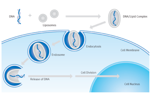
| − | + | [[Image:Sp16 M2D5 transfection schematic.png|thumb|right|500px|'''Overview of eukaryote cell transfection.''']]Before we go forward, let's take a moment to discuss how you will get the reporter DNA into the glioblastoma cells. In Module 1, we used transformation to move DNA into bacterial cells, here we'll complete a transfection with our eukaryotic cells. Mammalian cells can be transiently or stably transfected. For transient transfection, DNA is put into a cell and the transgene is expressed, but eventually the DNA is degraded and transgene expression is lost (''transgene'' is used to describe any gene that is introduced into a cell). For stable transfection, the DNA is introduced such that it is maintained indefinitely. Today you will be transiently transfecting your cultures of human fibroblast cells. | |
| − | + | There are several approaches that researchers have used to introduce DNA into a cell's nucleus. At one extreme there is ballistics. In essence, a small gun is used to shoot the DNA into the cell. This is both technically difficult and inefficient, and so we won't be using this approach! More common approaches are electroporation and lipofection. During electroporation, mammalian cells are mixed with DNA and subjected to a brief pulse of electrical current within a capacitor. The current causes the membranes (which are charged in a polar fashion) to momentarily flip around, making small holes in the cell membrane that the DNA can pass through. | |
| − | + | The most popular chemical approach for getting DNA into cells is called lipofection. With this technique, a DNA sample is coated with a special kind of lipid that is able to fuse with mammalian cell membranes. When the coated DNA is mixed with the cells, they engulf it through endocytosis. The DNA stays in the cytoplasm of the cell until the next cell division, at which time the cell’s nuclear membrane dissolves and the DNA has a chance to enter the nucleus. | |
| − | + | Tomorrow, 24 hours post-transfection, the teaching faculty will measure the fluorescent signal emitted from these cells. Because only cells that carry a repaired plasmid will fluoresce green, you can compare this information to the controls to calculate the NHEJ efficiency in reference to the specific variables you chose/choose. The first variable that you will consider is the type of DNA damage (''i.e.'' the DNA ends created by the restriction enzyme digest). The second variable is the effect of a drug reported to inhibit NHEJ repair in other model systems. The purpose for examining the role of drugs in NHEJ repair efficiency is two-fold: 1) targeting NHEJ inhibitors to cancer cells is important for the treatment of disease, and 2) chemical inhibitors are used widely in biological engineering, so it is useful to think about their strengths and limitations. | |
| + | |||
| + | Review the following inhibitors, then sign up on the [http://engineerbiology.org/wiki/Talk:20.109%28S16%29:Cell_preparation_for_DNA_repair_assays_%28Day5%29 M2D5 Discussion page] for the drug that you will use in your assay. Consider collaborating with another team to assess a single drug with two types of DNA damage...or one type of DNA damage with each of the drugs. You will use the drug you choose for two experiments, the flow cytometry-based DNA repair assay, and an irradiation-induced cell-death experiment. The latter is a 'kill curve' that will examine NHEJ inhibition with increasing doses of the drug following 4 Gy of irradiation to induce DNA damage. If the drug is effective, the DNA damage will not be repaired efficiently and the cells will not survive. | ||
<center> | <center> | ||
| Line 22: | Line 27: | ||
! Literature reference | ! Literature reference | ||
! Fun fact | ! Fun fact | ||
| − | |||
| − | |||
| − | |||
| − | |||
| − | |||
| − | |||
|- | |- | ||
| Loperamide hydrochloride | | Loperamide hydrochloride | ||
| − | | | + | | unknown (for NHEJ) |
| [http://www.scbt.com/datasheet-203116-loperamide-hydrochloride.html Santa Cruz] | | [http://www.scbt.com/datasheet-203116-loperamide-hydrochloride.html Santa Cruz] | ||
| − | | [[Media:M2D6paperS15.pdf | Goglia et al.]] | + | | [[Media:M2D6paperS15.pdf | Goglia ''et al.'']] |
| − | | | + | | also known as Imodium |
| − | + | ||
| − | + | ||
| − | + | ||
| − | + | ||
| − | + | ||
| − | + | ||
|- | |- | ||
|DMNB | |DMNB | ||
|DNA-PKcs inhibitor | |DNA-PKcs inhibitor | ||
|[http://www.scbt.com/datasheet-202142-dmnb.html Santa Cruz] | |[http://www.scbt.com/datasheet-202142-dmnb.html Santa Cruz] | ||
| − | |[http://www.ncbi.nlm.nih.gov/pubmed/14500812 Durant et al.] | + | |[http://www.ncbi.nlm.nih.gov/pubmed/14500812 Durant ''et al.''] |
| − | | | + | |chemical derivative of vanilla |
|} | |} | ||
</center> | </center> | ||
| − | == | + | ==Protocols== |
| − | + | Today the class will be divided between the two exercises below. | |
| − | + | ===Part 1: Transfection of cells with plasmid reporters=== | |
| − | + | '''Timing is important for this experiment, so complete all calculations and be sure of all manipulations before you begin.''' | |
| − | + | ||
| − | + | ||
| − | + | ||
| − | + | ||
| − | + | ||
| − | + | ||
| − | + | ||
| − | + | ||
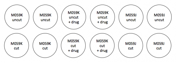
| − | + | In anticipation of your lipofection experiment, the teaching faculty plated 5,000 M059K or M059J cells per well in a 24-well dish ~48 h in advance of today's laboratory session. Review the plating schematic for your experiment that is shown below and be sure to note which cells are where! | |
| − | + | [[Image:Sp16 M2D5 plate map.png|thumb|center|600px|'''Lipofection plate map (top half of a 24-well plate).''' The top row will receive mixture A (see below, intact pMAX_EGFP_MCS), and the bottom row mixture B (see below, damaged pMAX_EGFP_MCS). Each condition will be prepared in duplicate.]] | |
| − | |||
| − | |||
| − | |||
| − | |||
| − | |||
| − | |||
| − | |||
| − | |||
| − | |||
| − | |||
| − | |||
| − | |||
<br style="clear:both" /> | <br style="clear:both" /> | ||
| − | Below is a | + | Below is a table to help you complete the calculations you need for your work in the tissue culture room. Please note the following procedural details before you work through the math: |
| − | + | *You will prepare enough of the 'LTX' solution for all of the wells represented in the plate map schematic above. To account for pipetting error, you will add an additional 10% of each component to the mixture. | |
| − | + | *You will prepare enough of the 'A' solution (containing intact pMAX_EGFP_MCS) for half of the wells represented in the plate map schematic above. To account for pipetting error, you will add an additional 10% of each component to the mixture. | |
| − | + | *You will prepare enough of the 'B' solution (containing damaged pMAX_EGFP_MCS) for half of the wells represented in the plate map schematic above. '''If you do not have enough damaged pMAX_EGFP_MCS from your preparation on M2D3, use the STOCK damaged pMAX_EGFP_MCS to supplement.''' To account for pipetting error, you will add an additional 10% of each component to the mixture. | |
| − | + | *Both the 'A' and 'B' solutions will include pMAX_mCherry as a transfection control. | |
| − | + | ||
| − | + | ||
| − | + | ||
| − | + | ||
| − | + | ||
| − | + | ||
| − | + | ||
| − | + | ||
| − | + | ||
| − | + | ||
| − | + | ||
| − | + | ||
| − | + | ||
| − | + | ||
| − | + | ||
<center> | <center> | ||
{| border="1" | {| border="1" | ||
| + | ! Tube | ||
! Component | ! Component | ||
! [Stock] ng/μL | ! [Stock] ng/μL | ||
| − | ! Amount per | + | ! Amount per well |
| − | ! | + | ! Total volume + pipetting error excess (μL) |
|- | |- | ||
| − | | LTX | + | | 'LTX' |
| + | | Lipofectamine | ||
| N/A | | N/A | ||
| − | | 0 | + | | 1.0 μL |
| − | | | + | | |
|- | |- | ||
| − | | | + | | |
| + | | OptiMEM | ||
| N/A | | N/A | ||
| − | | to total 25 μL | + | | to total 25 μL |
| − | | | + | | |
|- | |- | ||
| − | | | + | | 'A' |
| − | | | + | | pMAX_mCherry |
| − | | | + | | 250 ng/μL |
| − | | | + | | 250 ng |
| + | | | ||
|- | |- | ||
| − | | intact | + | | |
| − | | 250 | + | | intact pMAX_EGFP_MCS |
| − | | 250 | + | | 250 ng/μL |
| − | | | + | | 250 ng |
| + | | | ||
|- | |- | ||
| − | | damaged | + | | |
| − | | | + | | PLUS |
| − | | | + | | N/A |
| − | |250 | + | | 0.5 μL |
| + | | | ||
| + | |- | ||
| + | | | ||
| + | | OptiMEM | ||
| + | | N/A | ||
| + | | to total 25 μL | ||
| + | | | ||
| + | |- | ||
| + | | 'B' | ||
| + | | pMAX_mCherry | ||
| + | | 250 ng/μL | ||
| + | | 250 ng | ||
| + | | | ||
| + | |- | ||
| + | | | ||
| + | | YOUR damaged pMAX_EGFP_MCS | ||
| + | | Check your notes! | ||
| + | | 250 ng | ||
| + | | | ||
| + | |- | ||
| + | | | ||
| + | | STOCK ''PmeI''-cut pMAX_EGFP_MCS | ||
| + | | 100 ng/μL | ||
| + | | 250 ng | ||
| + | | | ||
| + | |- | ||
| + | | | ||
| + | | STOCK ''EcoRI''-''BglII''-cut pMAX_EGFP_MCS | ||
| + | | 150 ng/μL | ||
| + | | 250 ng | ||
| + | | | ||
|- | |- | ||
| − | | | + | | |
| − | | | + | | STOCK ''PstI-HF''-''BglII''-cut pMAX_EGFP_MCS |
| − | | | + | | 190 ng/μL |
| − | | | + | | 250 ng |
| + | | | ||
|- | |- | ||
| − | | PLUS | + | | |
| + | | PLUS | ||
| N/A | | N/A | ||
| − | | 0.5 | + | | 0.5 μL |
| − | | | + | | |
|- | |- | ||
| − | | | + | | |
| + | | OptiMEM | ||
| N/A | | N/A | ||
| − | | to total 25 μL | + | | to total 25 μL |
| − | | | + | | |
|- | |- | ||
|} | |} | ||
</center> | </center> | ||
| − | ===Part 2: Peer | + | Check with the teaching faculty to ensure your calculations are correct, then '''carefully read through the protocol and workflow schematic below...ask questions if any part of the exercise is unclear!''' When you are confident that you understand the steps necessary for the transfection, alert the teaching faculty that you are ready to begin your tissue culture work. |
| + | |||
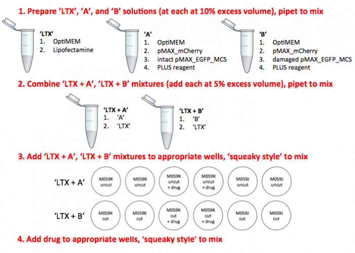
| + | '''Add all components in the order shown on the experimental overview schematic below and be sure to use the method listed.''' | ||
| + | |||
| + | #Pre-label all of the eppendorf tubes that you will need for the transfection procedure. | ||
| + | #Prepare the 'LTX' solution according to your calculations above. | ||
| + | #*The diluted LTX can sit at room temperature during the next two steps. | ||
| + | #Combine the OptiMEM and DNA for the 'A' and 'B' solutions, according to your calculations above. As a sanity check: | ||
| + | #*'A' is the control solution containing intact pMAX_EGFP_MCS | ||
| + | #*'B' is the experimental solution containing damaged pMAX_EGFP_MCS | ||
| + | #*Both solutions contain the transfection control pMAX_mCherry. | ||
| + | #Add the PLUS reagent to the 'A' and 'B' DNA solutions. | ||
| + | #Distribute the 'LTX' to 2 ''fresh'' eppendorf tubes, one labeled 'LTX + A' and one 'LTX + B'. Here include the volume needed for the appropriate number of wells plus 5% excess. | ||
| + | #*For example, if 50 μL were required, you would add 52.5 μL instead. | ||
| + | #Then add the 'A' solution (volume needed for the appropriate number of wells plus 5% excess) the 'LTX + A' tube containing from Step #5. Repeat for the 'B' solution. | ||
| + | #Incubate the mixtures from Step #6 for '''20 min''' in the tissue culture hood. | ||
| + | #During this incubation, give your cells fresh media: | ||
| + | #*Aspirate media from all 12 wells, making sure to use a different Pasteur pipet for M059K and M059J cells. | ||
| + | #*Add 500 μL (using the P1000) of fresh pre-warmed media to each well. | ||
| + | #Add 50 μL of the appropriate 'LTX + A' and 'LTX + B' mixtures to each well according to the plate map above. '''Read through the following tips before proceeding:''' | ||
| + | #*Add the 'LTX + A' and LTX + B' mixtures drop-by-drop while making a circle with your pipet in the well, then immediately rock the plate back-and-forth two times to distribute the mixture within the well. | ||
| + | #*Change tips between every well. | ||
| + | #*After distributing to all 12 wells, do an additional mixing step for the whole plate, 'squeaky style': a few each of horizontal and vertical tilts. | ||
| + | #Lastly, you will add 2 μL of the drug you selected to the appropriate wells. | ||
| + | #*Be sure to complete another squeaky style mixing step for the whole plate. | ||
| + | #Label your 24-well plate with the date, your team color and sample information then move it to the incubator. | ||
| + | |||
| + | |||
| + | [[Image:Sp16 M2D5 experimental overview.png|thumb|center|700px|'''Lipofection experimental overview and workflow.''']] | ||
| + | |||
| + | ===Part 2: Peer review of Methods homework=== | ||
Now that you've had two rounds of feedback and three rounds of drafting practice, you will put your knowledge to work by reviewing and correcting the Methods sections of your peers. First, read the text below as a guide to what your instructors look for while grading Methods sections. Next, read completely through the text of Methods draft that you will offer comments on. Finally, using whatever style is easiest for you, offer specific comments to your fellow 109er about what they have done well and what needs further work. Complete this activity using the "golden rule" -- offer the type of feedback that you hope to also receive. One of the best ways to improve your own skills is to teach something, so take advantage of this opportunity. | Now that you've had two rounds of feedback and three rounds of drafting practice, you will put your knowledge to work by reviewing and correcting the Methods sections of your peers. First, read the text below as a guide to what your instructors look for while grading Methods sections. Next, read completely through the text of Methods draft that you will offer comments on. Finally, using whatever style is easiest for you, offer specific comments to your fellow 109er about what they have done well and what needs further work. Complete this activity using the "golden rule" -- offer the type of feedback that you hope to also receive. One of the best ways to improve your own skills is to teach something, so take advantage of this opportunity. | ||
| − | Once you have completed your review, please turn in your comments and the Methods section that you reviewed to the front bench. | + | Once you have completed your review, please turn in your comments and the Methods section that you reviewed to the front bench. Leslie and Maxine will provide further comments on the Methods section that you reviewed, as well as comments to you as the reviewer to help you with these activities in the future. |
====(A) Holistic view of Methods section==== | ====(A) Holistic view of Methods section==== | ||
| Line 166: | Line 193: | ||
#'''Descriptive sub-section titles''' | #'''Descriptive sub-section titles''' | ||
#*Do the sub-section titles give enough information for the audience to easily pinpoint where the information they need will be found? | #*Do the sub-section titles give enough information for the audience to easily pinpoint where the information they need will be found? | ||
| − | #*Are the titles specific (i.e. Culture of | + | #*Are the titles specific (''i.e.'' Culture of M059K and M059J cells) or could they belong in any paper that involves cell culture (''i.e.'' Cell culture)? |
#'''Introductory sentences''' | #'''Introductory sentences''' | ||
| − | #*Does each sub-section start with an introductory sentence that states the goal of the experiment that was done? (For example, A Western blot was completed to determine | + | #*Does each sub-section start with an introductory sentence that states the goal of the experiment that was done? (For example, A Western blot was completed to determine DNA-PK expression in M059K and M059J cell lines.) |
====(B) Specific experimental details==== | ====(B) Specific experimental details==== | ||
| Line 201: | Line 228: | ||
#*Is the section easy to read or are there words missing/misspelled/misused? | #*Is the section easy to read or are there words missing/misspelled/misused? | ||
| − | For this activity, please comment on each of the three major evaluation criteria (A-C) listed above. | + | For this activity, please comment on each of the three major evaluation criteria (A-C) listed above. Do this by following the format that your teaching faculty employ -- numbering the paper and then writing an accompanying document. You will submit your comments at the start of your laboratory section on M2D6. |
| − | + | ||
| − | + | ||
| − | + | ||
| − | + | ||
| − | + | ||
| − | + | ||
| − | + | ||
| − | + | ||
| − | + | ||
| − | + | ||
| − | + | ||
| − | + | ||
| − | + | ||
==Reagent list== | ==Reagent list== | ||
| − | + | *Lipofectamine® LTX Reagent with PLUS™ Reagent from Life Technologies | |
| − | *Lipofectamine® LTX Reagent with PLUS™ Reagent | + | *Opti-MEM reduced serum medium from Life Technologies |
| − | *Opti-MEM reduced serum medium | + | *Loperamide hydrochloride, 2.8 μM |
| + | *DMNB (4,5-dimethoxy-2-nitrobenzaldehyde), 15 μM | ||
==Navigation links== | ==Navigation links== | ||
| − | Next | + | Next day: [[20.109(S16):DNA repair assays (Day6)| DNA repair assays]] |
| − | Previous | + | Previous day: [[20.109(S16):Journal Club I (Day4)| Journal Club I]] |
Latest revision as of 18:34, 28 March 2016
Contents
Introduction
Today you will (finally) use the pMAX_EGFP_MCS reporter construct you prepared and purified during the previous laboratory session with the human glioblastoma cell lines to assess the NHEJ mechanism. Remember that the M059K cells are the wild-type cells, which carry DNA-PKcs, and the M059J cells are DNA-PKcs mutants. This suggests that the M059J cells are unable to perform NHEJ and will, therefore, be the negative control for your experiments. In your experimental setup today you will transfect M059K cells with damaged pMAX_EGFP_MCS and you will also transfect cells with intact, or undamaged, pMAX_EGFP_MCS. The cells transfected with the damaged reporter will be the experimental samples and those transfected with the intact reporter will be used as the positive controls. Let's take a closer look:
- Negative control: The M059J cells are unable to perform NHEJ and therefore cells transfected with damaged reporter will emit background fluorescence and allow you to visualize the level of repair that occurs via another pathway, therefore it is used to show when the NHEJ efficiency is equal to 0%.
- Positive control: The subset of M059K cells that are transfected with intact reporter will show the frequency of fluorescent signal when the NHEJ efficiency is equal to 100%.
- Experimental samples: These are the cells that will give you data!
There are several approaches that researchers have used to introduce DNA into a cell's nucleus. At one extreme there is ballistics. In essence, a small gun is used to shoot the DNA into the cell. This is both technically difficult and inefficient, and so we won't be using this approach! More common approaches are electroporation and lipofection. During electroporation, mammalian cells are mixed with DNA and subjected to a brief pulse of electrical current within a capacitor. The current causes the membranes (which are charged in a polar fashion) to momentarily flip around, making small holes in the cell membrane that the DNA can pass through.
The most popular chemical approach for getting DNA into cells is called lipofection. With this technique, a DNA sample is coated with a special kind of lipid that is able to fuse with mammalian cell membranes. When the coated DNA is mixed with the cells, they engulf it through endocytosis. The DNA stays in the cytoplasm of the cell until the next cell division, at which time the cell’s nuclear membrane dissolves and the DNA has a chance to enter the nucleus.
Tomorrow, 24 hours post-transfection, the teaching faculty will measure the fluorescent signal emitted from these cells. Because only cells that carry a repaired plasmid will fluoresce green, you can compare this information to the controls to calculate the NHEJ efficiency in reference to the specific variables you chose/choose. The first variable that you will consider is the type of DNA damage (i.e. the DNA ends created by the restriction enzyme digest). The second variable is the effect of a drug reported to inhibit NHEJ repair in other model systems. The purpose for examining the role of drugs in NHEJ repair efficiency is two-fold: 1) targeting NHEJ inhibitors to cancer cells is important for the treatment of disease, and 2) chemical inhibitors are used widely in biological engineering, so it is useful to think about their strengths and limitations.
Review the following inhibitors, then sign up on the M2D5 Discussion page for the drug that you will use in your assay. Consider collaborating with another team to assess a single drug with two types of DNA damage...or one type of DNA damage with each of the drugs. You will use the drug you choose for two experiments, the flow cytometry-based DNA repair assay, and an irradiation-induced cell-death experiment. The latter is a 'kill curve' that will examine NHEJ inhibition with increasing doses of the drug following 4 Gy of irradiation to induce DNA damage. If the drug is effective, the DNA damage will not be repaired efficiently and the cells will not survive.
| Drug | Mechanism of action | Vendor website | Literature reference | Fun fact |
|---|---|---|---|---|
| Loperamide hydrochloride | unknown (for NHEJ) | Santa Cruz | Goglia et al. | also known as Imodium |
| DMNB | DNA-PKcs inhibitor | Santa Cruz | Durant et al. | chemical derivative of vanilla |
Protocols
Today the class will be divided between the two exercises below.
Part 1: Transfection of cells with plasmid reporters
Timing is important for this experiment, so complete all calculations and be sure of all manipulations before you begin.
In anticipation of your lipofection experiment, the teaching faculty plated 5,000 M059K or M059J cells per well in a 24-well dish ~48 h in advance of today's laboratory session. Review the plating schematic for your experiment that is shown below and be sure to note which cells are where!
Below is a table to help you complete the calculations you need for your work in the tissue culture room. Please note the following procedural details before you work through the math:
- You will prepare enough of the 'LTX' solution for all of the wells represented in the plate map schematic above. To account for pipetting error, you will add an additional 10% of each component to the mixture.
- You will prepare enough of the 'A' solution (containing intact pMAX_EGFP_MCS) for half of the wells represented in the plate map schematic above. To account for pipetting error, you will add an additional 10% of each component to the mixture.
- You will prepare enough of the 'B' solution (containing damaged pMAX_EGFP_MCS) for half of the wells represented in the plate map schematic above. If you do not have enough damaged pMAX_EGFP_MCS from your preparation on M2D3, use the STOCK damaged pMAX_EGFP_MCS to supplement. To account for pipetting error, you will add an additional 10% of each component to the mixture.
- Both the 'A' and 'B' solutions will include pMAX_mCherry as a transfection control.
| Tube | Component | [Stock] ng/μL | Amount per well | Total volume + pipetting error excess (μL) |
|---|---|---|---|---|
| 'LTX' | Lipofectamine | N/A | 1.0 μL | |
| OptiMEM | N/A | to total 25 μL | ||
| 'A' | pMAX_mCherry | 250 ng/μL | 250 ng | |
| intact pMAX_EGFP_MCS | 250 ng/μL | 250 ng | ||
| PLUS | N/A | 0.5 μL | ||
| OptiMEM | N/A | to total 25 μL | ||
| 'B' | pMAX_mCherry | 250 ng/μL | 250 ng | |
| YOUR damaged pMAX_EGFP_MCS | Check your notes! | 250 ng | ||
| STOCK PmeI-cut pMAX_EGFP_MCS | 100 ng/μL | 250 ng | ||
| STOCK EcoRI-BglII-cut pMAX_EGFP_MCS | 150 ng/μL | 250 ng | ||
| STOCK PstI-HF-BglII-cut pMAX_EGFP_MCS | 190 ng/μL | 250 ng | ||
| PLUS | N/A | 0.5 μL | ||
| OptiMEM | N/A | to total 25 μL |
Check with the teaching faculty to ensure your calculations are correct, then carefully read through the protocol and workflow schematic below...ask questions if any part of the exercise is unclear! When you are confident that you understand the steps necessary for the transfection, alert the teaching faculty that you are ready to begin your tissue culture work.
Add all components in the order shown on the experimental overview schematic below and be sure to use the method listed.
- Pre-label all of the eppendorf tubes that you will need for the transfection procedure.
- Prepare the 'LTX' solution according to your calculations above.
- The diluted LTX can sit at room temperature during the next two steps.
- Combine the OptiMEM and DNA for the 'A' and 'B' solutions, according to your calculations above. As a sanity check:
- 'A' is the control solution containing intact pMAX_EGFP_MCS
- 'B' is the experimental solution containing damaged pMAX_EGFP_MCS
- Both solutions contain the transfection control pMAX_mCherry.
- Add the PLUS reagent to the 'A' and 'B' DNA solutions.
- Distribute the 'LTX' to 2 fresh eppendorf tubes, one labeled 'LTX + A' and one 'LTX + B'. Here include the volume needed for the appropriate number of wells plus 5% excess.
- For example, if 50 μL were required, you would add 52.5 μL instead.
- Then add the 'A' solution (volume needed for the appropriate number of wells plus 5% excess) the 'LTX + A' tube containing from Step #5. Repeat for the 'B' solution.
- Incubate the mixtures from Step #6 for 20 min in the tissue culture hood.
- During this incubation, give your cells fresh media:
- Aspirate media from all 12 wells, making sure to use a different Pasteur pipet for M059K and M059J cells.
- Add 500 μL (using the P1000) of fresh pre-warmed media to each well.
- Add 50 μL of the appropriate 'LTX + A' and 'LTX + B' mixtures to each well according to the plate map above. Read through the following tips before proceeding:
- Add the 'LTX + A' and LTX + B' mixtures drop-by-drop while making a circle with your pipet in the well, then immediately rock the plate back-and-forth two times to distribute the mixture within the well.
- Change tips between every well.
- After distributing to all 12 wells, do an additional mixing step for the whole plate, 'squeaky style': a few each of horizontal and vertical tilts.
- Lastly, you will add 2 μL of the drug you selected to the appropriate wells.
- Be sure to complete another squeaky style mixing step for the whole plate.
- Label your 24-well plate with the date, your team color and sample information then move it to the incubator.
Part 2: Peer review of Methods homework
Now that you've had two rounds of feedback and three rounds of drafting practice, you will put your knowledge to work by reviewing and correcting the Methods sections of your peers. First, read the text below as a guide to what your instructors look for while grading Methods sections. Next, read completely through the text of Methods draft that you will offer comments on. Finally, using whatever style is easiest for you, offer specific comments to your fellow 109er about what they have done well and what needs further work. Complete this activity using the "golden rule" -- offer the type of feedback that you hope to also receive. One of the best ways to improve your own skills is to teach something, so take advantage of this opportunity.
Once you have completed your review, please turn in your comments and the Methods section that you reviewed to the front bench. Leslie and Maxine will provide further comments on the Methods section that you reviewed, as well as comments to you as the reviewer to help you with these activities in the future.
(A) Holistic view of Methods section
When the instructors first read your Methods section, we begin by taking a holistic view of its structure. There are three main elements that all good methods sections must contain:
- An appropriate number of sub-sections
- Has the author divided the Methods into logical sub-sections based upon an entire experiment and not based upon completion day in the 20.109 lab?
- Descriptive sub-section titles
- Do the sub-section titles give enough information for the audience to easily pinpoint where the information they need will be found?
- Are the titles specific (i.e. Culture of M059K and M059J cells) or could they belong in any paper that involves cell culture (i.e. Cell culture)?
- Introductory sentences
- Does each sub-section start with an introductory sentence that states the goal of the experiment that was done? (For example, A Western blot was completed to determine DNA-PK expression in M059K and M059J cell lines.)
(B) Specific experimental details
Next, we examine each Methods section to make sure that all of the details that are required to replicate your study are included. For this assignment, the important details are as follows (here we've included them within the sub-sections that would be appropriate for M2D1-D3):
- Cell culture sub-section
- cell types and origin (where did you get them?)
- cell culture media formulation
- culture conditions (temperature, CO2%, and relative humidity)
- Note: later you may choose to include the inhibitor name, concentration, and treatment times in this section.
- Western blot sub-section
- cell seeding density and incubation time
- lysis buffer formulation and lysis conditions (PBS rinse, temperature)
- protein quantification reagent, total protein separated on gel
- SDS-PAGE gel %, TE buffer formulation, electrophoresis conditions
- transfer buffer formulation, transfer conditions, membrane type, blocking buffer
- names of antibodies and dilutions, incubation times
- washing conditions and blot development
- DNA damage sub-section
- name of plasmid, mass digested
- name of enzymes, concentration of enzyme, reaction buffer
- reaction conditions, evaluation by electrophoresis
(C) Quality of writing
Finally, we provide comments on the writing style employed in each Methods section, concentrating on the following:
- Is this a protocol or a formal Methods section?
- Are volumes, vortexing steps, incubation times, etc. that are extraneous (or flexible) included? If so, this is a protocol.
- Is it concise?
- Are "filler words" avoided? Are sentences combined where possible to eliminate extraneous phrases and/or information?
- Is it logical?
- Do the 'steps' follow a logical progression? Again, is the section constructed to explain an experiment as a whole and not simply listed in the order in which it was performed in class?
- Is it written in complete sentences?
- Is the section easy to read or are there words missing/misspelled/misused?
For this activity, please comment on each of the three major evaluation criteria (A-C) listed above. Do this by following the format that your teaching faculty employ -- numbering the paper and then writing an accompanying document. You will submit your comments at the start of your laboratory section on M2D6.
Reagent list
- Lipofectamine® LTX Reagent with PLUS™ Reagent from Life Technologies
- Opti-MEM reduced serum medium from Life Technologies
- Loperamide hydrochloride, 2.8 μM
- DMNB (4,5-dimethoxy-2-nitrobenzaldehyde), 15 μM
Next day: DNA repair assays
Previous day: Journal Club I


